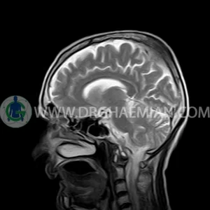
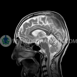
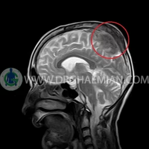
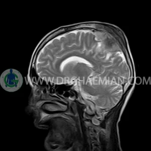
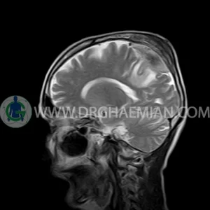
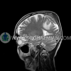
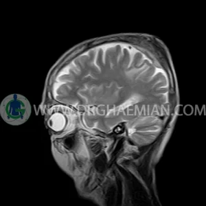
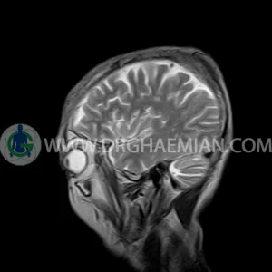
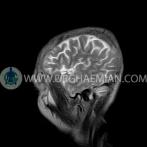
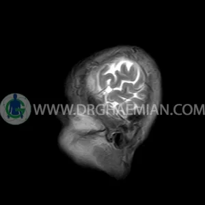
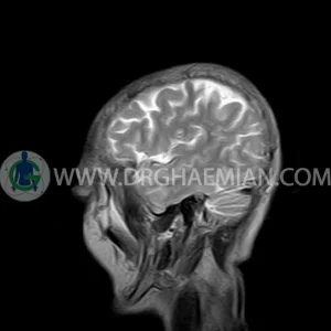
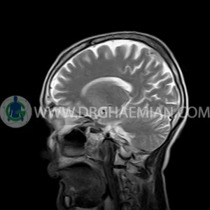
ام آر آی مغز با استفاده آهنربا های قوی و امواج رادیویی تصاویری از مغز و بافت های عصبی پیرامونی آن ایجاد می کند. در این کیس مننژیوم به همراه هیپراستوز استخوانی سمت چپ دیده می شود.


گزارش پزشک
BRAIN MRI
(Without contrast)
Technique:Axial FLAIR, Axial, sagittal, FSE T2, coronal T1 .
REPORT :
The interhemispheric fissure is centered on the midline .
No abnormalities are seen in the basal ganglia, int. capsule, corpus callosum, Thalamus and tectal plate.
The sella and pituitary – pineal g .are normal and parasellar, suprasellar structures are unremarkable.
The cerebello pontine angle area appears normal on each side.
The internal acoustic meatus has normal width .
Major intracranial vascular structures, pericavernous spaces and visualized intracranial nerve complex are normal.
The paranasal sinuses are clear and aerated with no evidence of bone erosion or destruction, fluid collection, cystic retention and mucosal thickening.
a well – defined dural based mass lesion ( 27x55mm ) in left parietal region with signal change in adajacent bone suggestive for meningioma with bone hyperosteosis
mass effect & edema in left parietal lobe
are seen
COMMENT : MRI with contrast is recommended .
