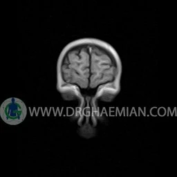
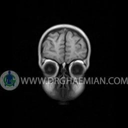
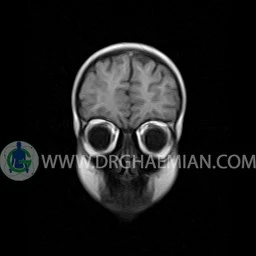
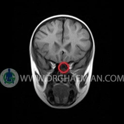
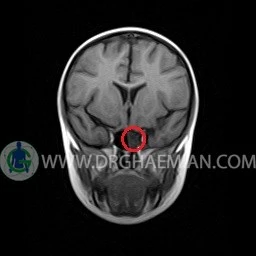
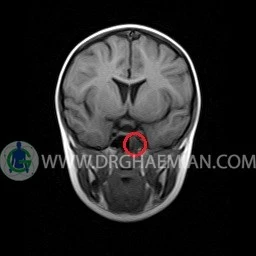
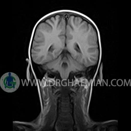
ام آر آی مغز با استفاده آهنربا های قوی و امواج رادیویی تصاویری از مغز و بافت های عصبی پیرامونی آن ایجاد می کند. در این کیس سینوزیت حاد سمت چپ به همراه ماستوئیدیت دیده می شود.


گزارش پزشک
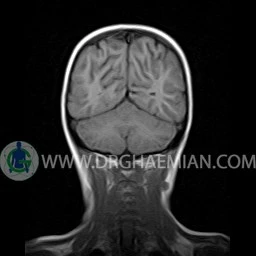
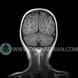
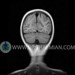
BRAIN MRI
(With & Without contrast)
Technique : Axial FLAIR, Axial, sagittal, FSE T2, coronal T1 & post contrast Axial-sagittal – coronal T1.
REPORT :
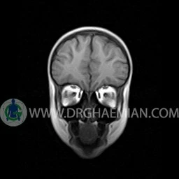
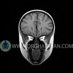
The interhemispheric fissure is centered on the midline .
The cerebral ventricles / cisterns are normal size and shape .
The cortex and white matter show normal signal intensity .
No abnormalities are seen in the basal ganglia, int. capsule, corpus callosum, Thalamus and tectal plate.
The brain stem, cerebellum show no abnormal changes in signal characteristics.
The sella and pituitary – pineal g .are normal and parasellar, suprasellar structures are unremarkable.
The orbital contents, falx, dura and calvaria are unremarkable .
Major intracranial vascular structures, pericavernous spaces and visualized intracranial nerve complex are normal .
There is no evidence of abnormal enhancement within cranium .
– Mucosal thickening with fluid level in left mastoidal cells suggestive for mastoiditis
– Mucosal thickening with fluid in left sphenoid sinus suggestive for acute sinusitis
are seen
