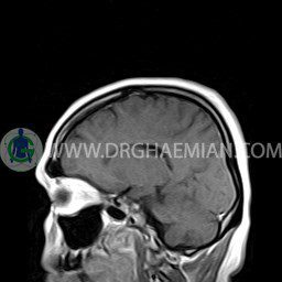
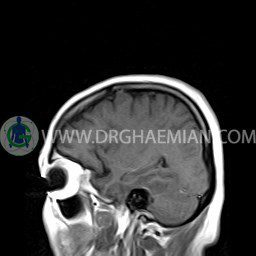
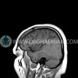
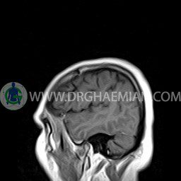
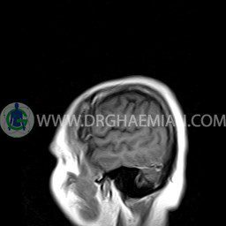
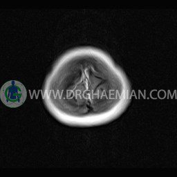
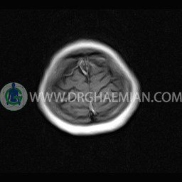
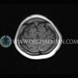
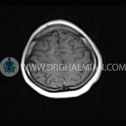
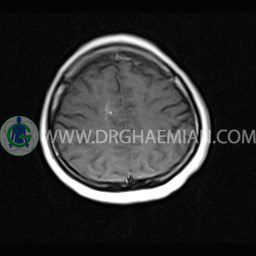
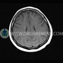
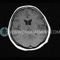
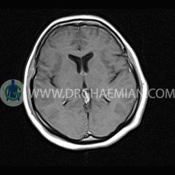
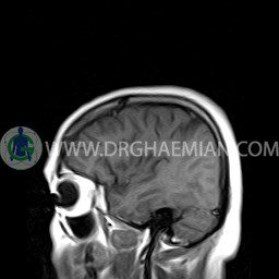
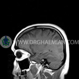
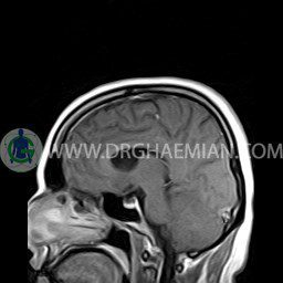
ام آر آی مغز یک روش تصویربرداری است که با استفاده آهنربا های قوی و امواج رادیویی تصاویری از مغز و بافت های عصبی پیرامونی آن ایجاد می کند. در این کیس آنژیوم عنکبوتی و سلای نسبتا خالی دیده می شود.


گزارش پزشک :
BRAIN MRI
(With contrast & comprassion with previous without contrast MRI -1401/08/29)
Technique: Axial T1 & post contrast axial – sagital – coronal T1 .
coronal T1 & post contrast Axial-sagittal – coronal T1 . (with)
REPORT :
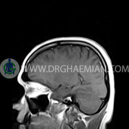
The interhemispheric fissure is centered on the midline .
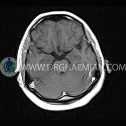
No abnormalities are seen in the basal ganglia , int . capsule, corpus callosum , Thalamus and tectal plate .
The brain stem , cerebellum show no abnormal changes in signal characteristics.
The cerebello pontine angle area appears normal on each side.
The internal acoustic meatus has normal width .
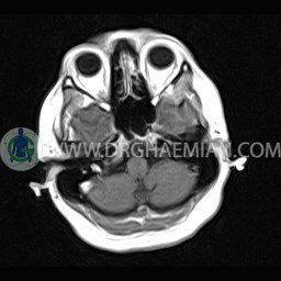
The orbital contents , falx , dura and calvaria are unremarkable .
Major intracranial vascular structures , pericavernous spaces and visualized intracranial nerve complex are normal .
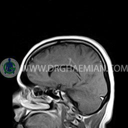
The paranasal sinuses are clear and aerated with no evidence of bone erosion or destruction, fluid collection , cystic retention and mucosal thickening.
– Dilated vein with palm tree around them in right centrum semovale suggestive for spider angioma ( develpmental venous anomaly )
– Small foci of white matter hyperintensity (deep W.M. ischemia- Fazekas 1)
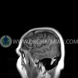
– Extension of suprasella cistern to sella with thin pituitary gland in floor of sella (partial empty sella)
are seen.
– gliosis & hemorrhagic changes is not seen .
