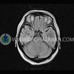
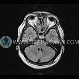
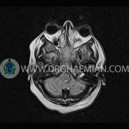
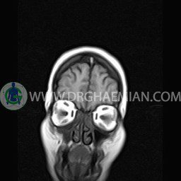
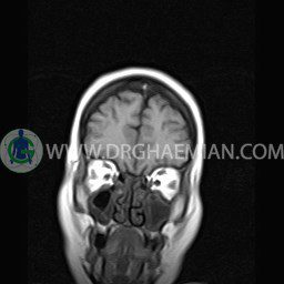
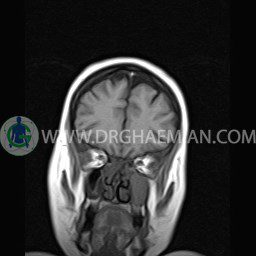
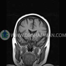
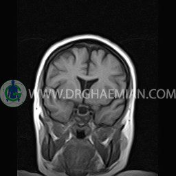
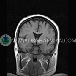
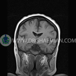
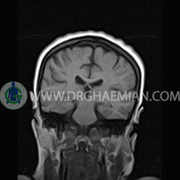
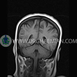
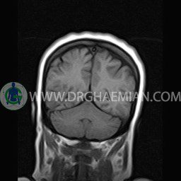
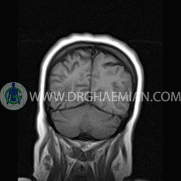
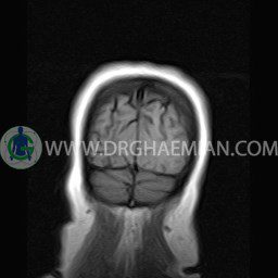
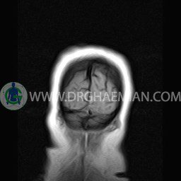
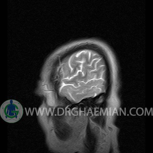
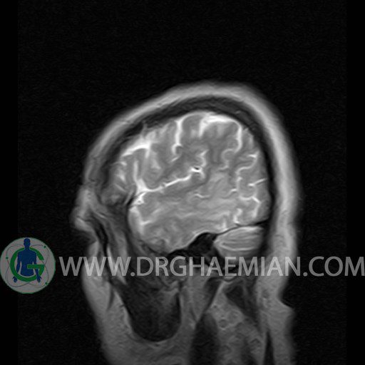
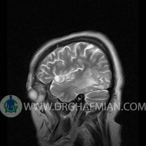
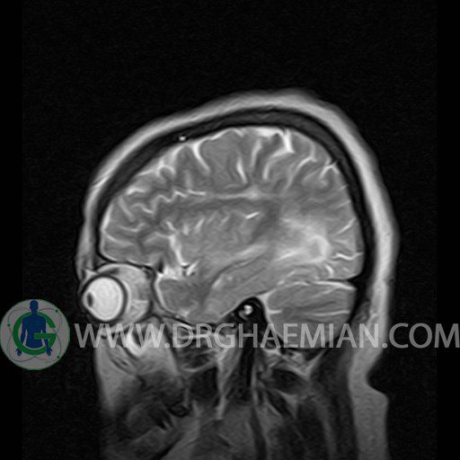
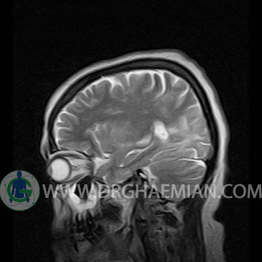
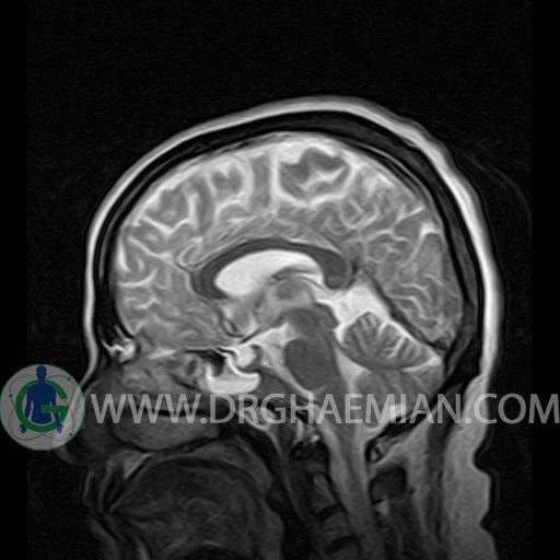
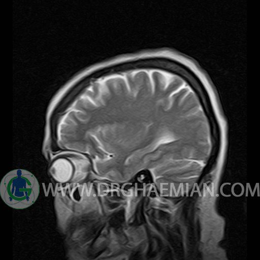
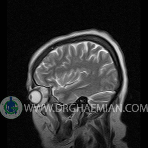
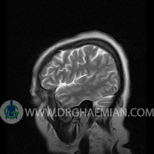
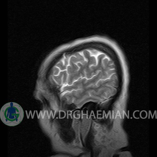
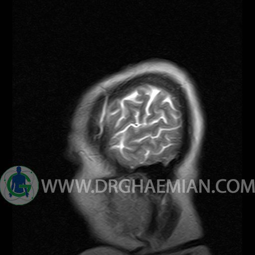
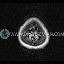
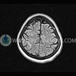
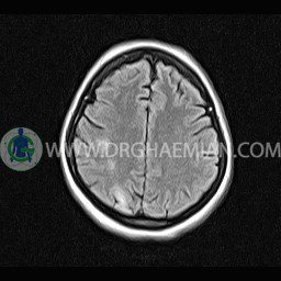
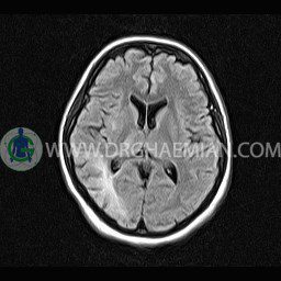
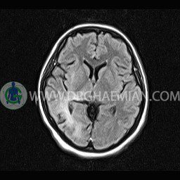
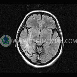
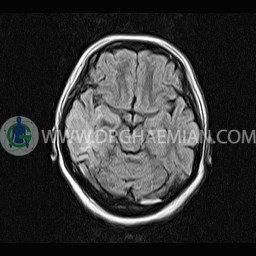
ام آر آی مغز یک روش تصویربرداری است که با استفاده آهنربا های قوی و امواج رادیویی تصاویری از مغز و بافت های عصبی پیرامونی آن ایجاد می کند. در این کیس سربریت و تغییرات اسکمیک، سلای خالی و ذخیم شدن مخاط در سینوس های پارانیزال دیده می شود.


گزارش پزشک :
BRAIN MRI
(Without contrast)
Technique:Axial FLAIR , Axial , sagittal , FSE T2 , coronal T1 .
REPORT :
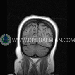
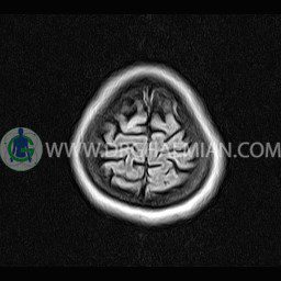
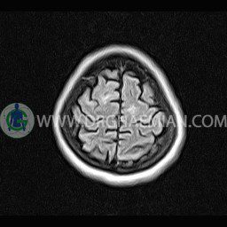
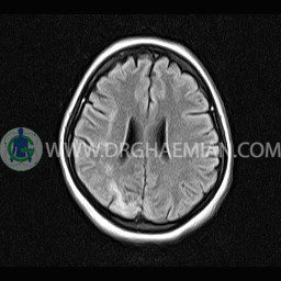
The interhemispheric fissure is centered on the midline .
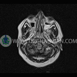
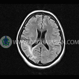
The cerebral ventricles / cisterns are normal size and shape .
The brain stem , cerebellum show no abnormal changes in signal characteristics.
The cerebello pontine angle area appears normal on each side.
The internal acoustic meatus has normal width .
The orbital contents , falx , dura and calvaria are unremarkable .
Major intracranial vascular structures , pericavernous spaces and visualized intracranial nerve complex are normal .
– Hyperintensity in subcortical and periventricular white matter of right parietotemporal association and in right thalamus suggestive for ischemic changes & cerebritis
– Multifocal hyperintensity in right & left centrum semiovale suggestive for ischemic changes
– Extension of suprasella cistern to sella with thin pituitary gland in floor of sella (empty sella)
– Mucosal thickening in paranasal sinuses with air-fluid level in maxillary sinuses
are seen.
COMMENT: Clinical correlation and MRI with contrast are recommended
