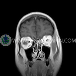
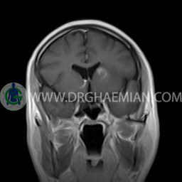
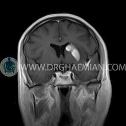
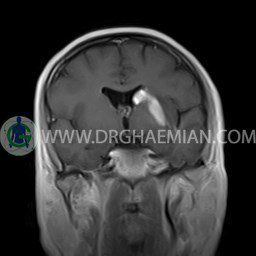
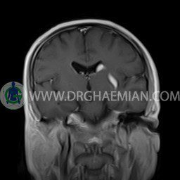
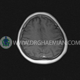
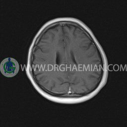
ام آر آی مغز یک روش تصویربرداری است که با استفاده از آهنربا های قوی و امواج رادیویی تصاویری از مغز و بافت های عصبی پیرامونی آن ایجاد می کند. در این کیس گلیوم پوتامن همراه با متاستاز مغز دیده می شود.
گزارش پزشک :
BRAIN MRI
(With & Without contrast)
Technique:Axial FLAIR , Axial , sagittal , FSE T2 , coronal T1 & post contrast Axial-sagittal – coronal T1 .
REPORT :
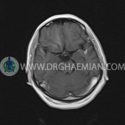
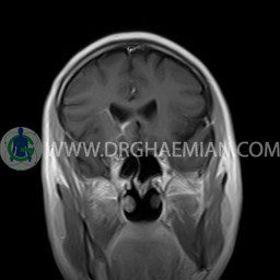
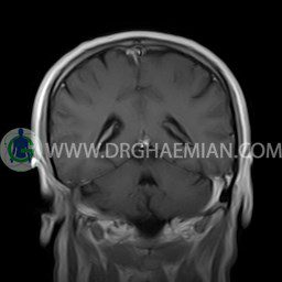
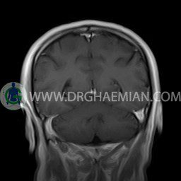
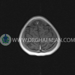
The interhemispheric fissure is centered on the midline .
The sella and pituitary – pineal g .are normal and parasellar, suprasellar structures are unremarkable.
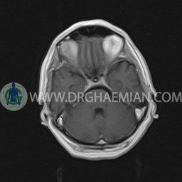
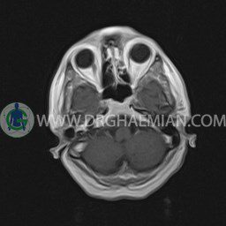
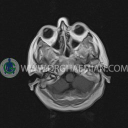
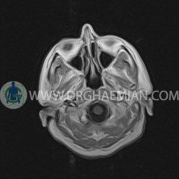
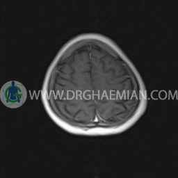
The cerebello pontine angle area appears normal on each side.
The internal acoustic meatus has normal width .
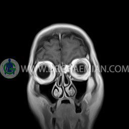
The orbital contents , falx , dura and calvaria are unremarkable .
Major intracranial vascular structures , pericavernous spaces and visualized intracranial nerve complex are normal .
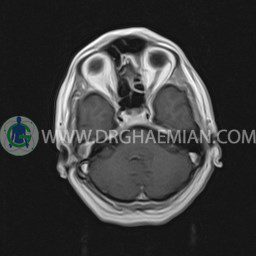
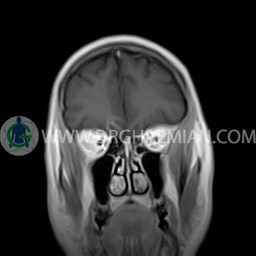
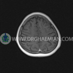
The paranasal sinuses are clear and aerated with no evidence of bone erosion or destruction, fluid collection , cystic retention and mucosal thickening.
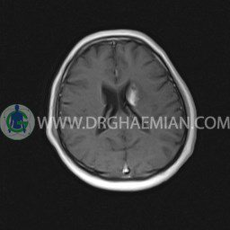
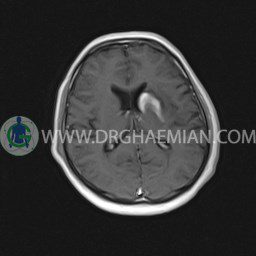
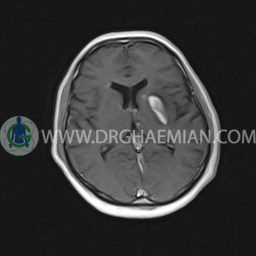
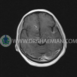
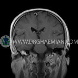
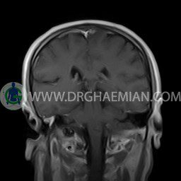
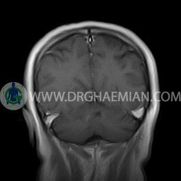
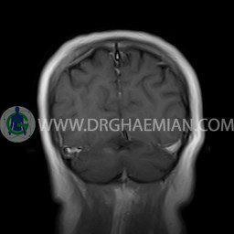
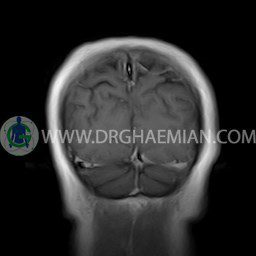
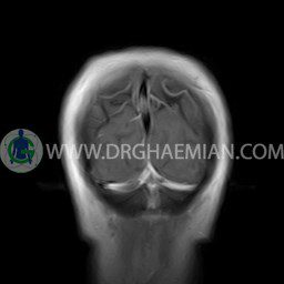
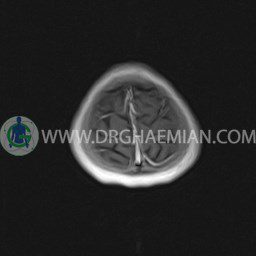
– A well – defined mass lesion ( 25 x 35 mm ) in left putamen- caudate nucleus ( high T2 – FLAIR & low T1 ) with mass effect on left lateral ventricle & with post contrast bright enhancement
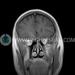
– Focal signal change at inferomedial of right frontal lobe & in left temporal pole ( high T2 – FLAIR ) with post contrast gyriform enhancement suggestive for glioma of putamen – caudate nucleus with cerebral matstasis in right frontal & left temporal lobes
are seen
