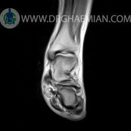
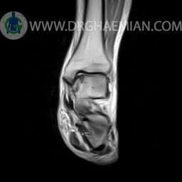
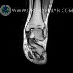
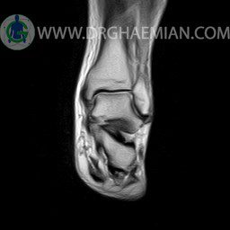
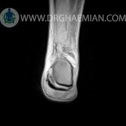
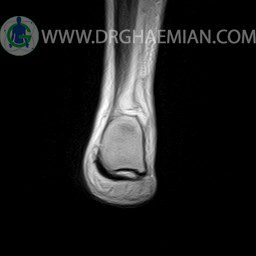
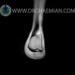
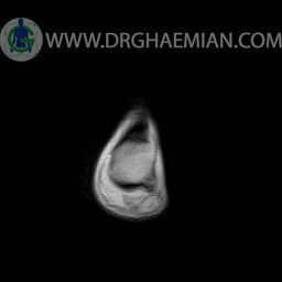
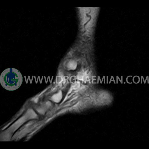
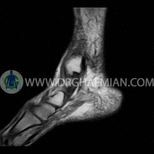
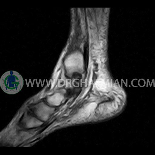
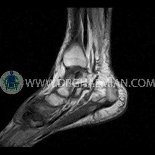
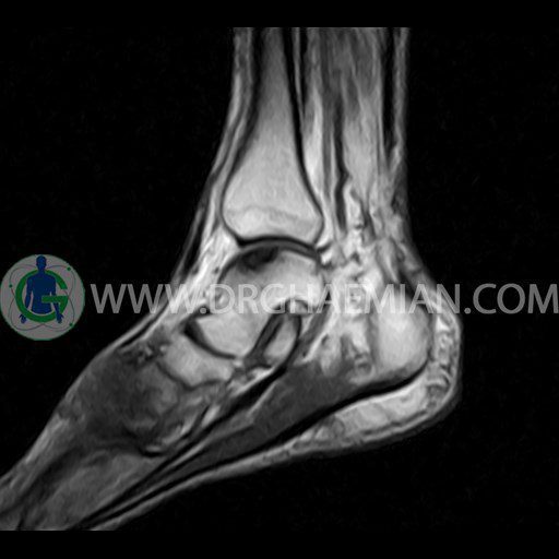
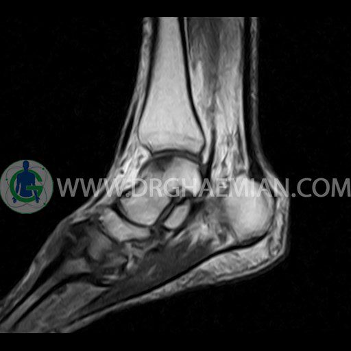
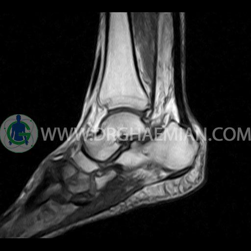
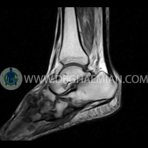
پزشکان اغلب از تصویربرداری ام آر آی برای تشخیص و درمان عارضه های پزشکی که فقط با استفاده از اشعه ایکس یا میدان مغناطیسی و امواج رادیویی قابل مشاهده است، استفاده می کنند. دستگاه ام آر آی تصاویر دقیق از ساختار های داخلی بدن ایجاد می کند. در این کیس ضایعه استئوکندرال مچ پا، تورم نسج نرم، تغییرات DJD، سینوویت گذرا و تنوسینوویت دیده می شود.
گزارش پزشک :
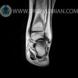
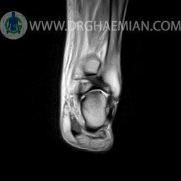
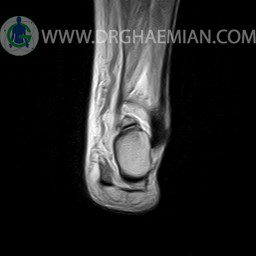
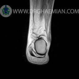
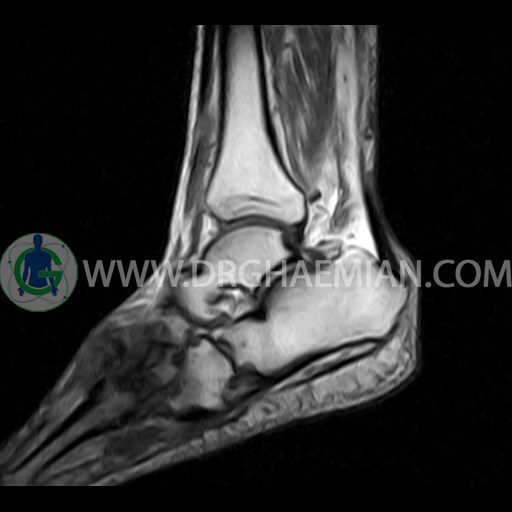
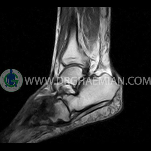
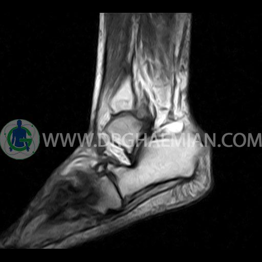
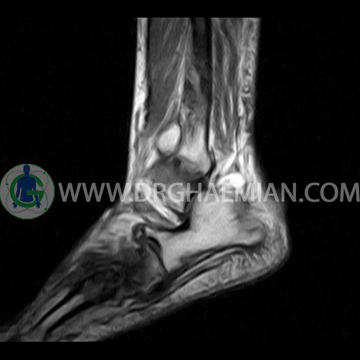
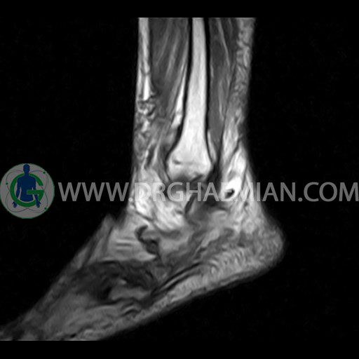
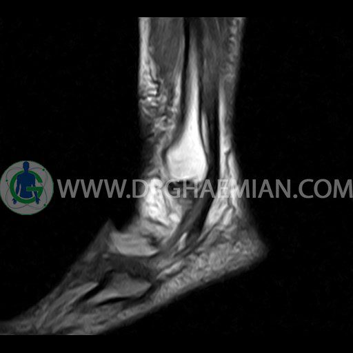
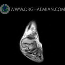
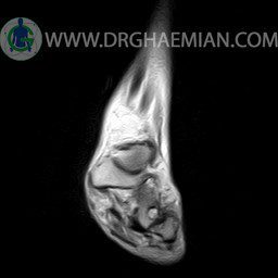
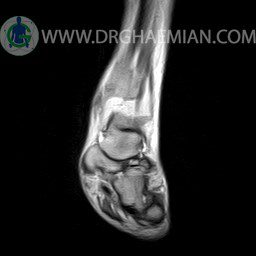
LEFT ANKLE MRI
(Without contrast)
Technique :Axial T2 , coronal , sagittal T1 and T2 ,sagittal T2 fat sat .
REPORT:
The bones comprising the ankle joint show normal position and configuration.
The bone marrow signal is normal.
The joint space is of normal width.
The cortex shows normal thickness and smooth contours , especially along the tibiotalar articular surfaces.
There are no subchondral signal changes and no osteophytes.
The lateral and medial ligaments are normal in their course , width and signal characteristics.
The talocalcaneal and talonavicular joints appear normal.
The achilles tendon is normal in its course , width and signal characteristics and the preachilles fat is clear.
The ankle tendons , ligaments and plantar aponeurosis are unremarkable.
Tarsal sinus is normal in shape and signal intensity.
-osteochondral lesion at medial of tallar dom with articular surface irregularity
– Soft tissue swelling around the ankle
– DJD changes in tarso metatarsal joint , in first , second & third fingers
– Tibio – talo – calcaneus joint effusion suggestive for transient synovitis
– Fluid around the medial tendon suggestive for tenosynovitis
are seen
