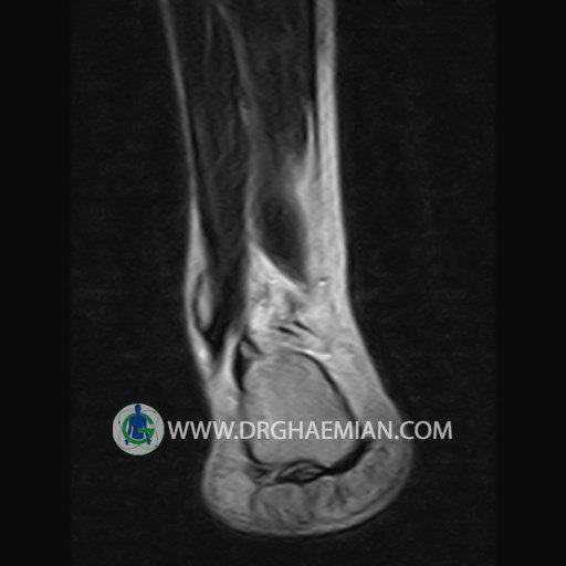
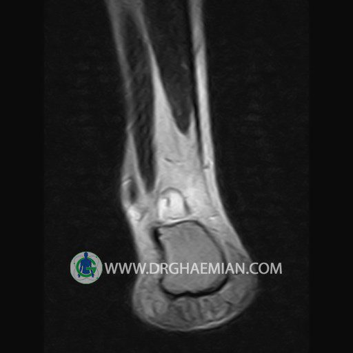
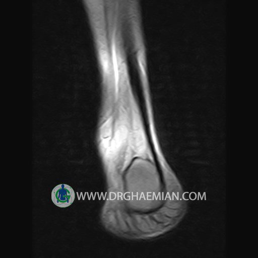
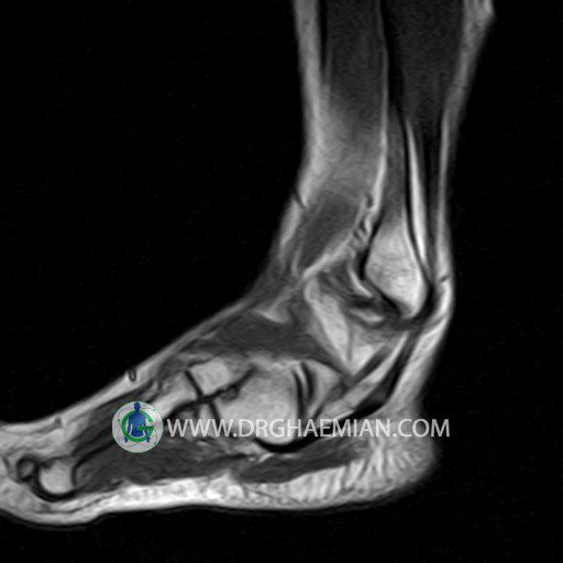
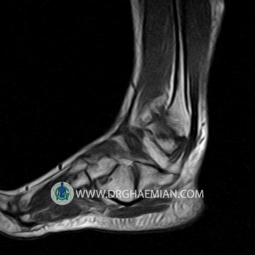
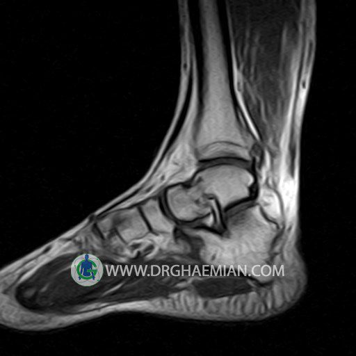
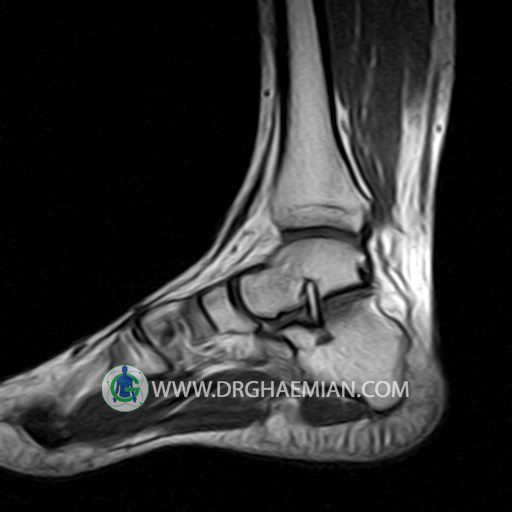
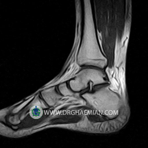
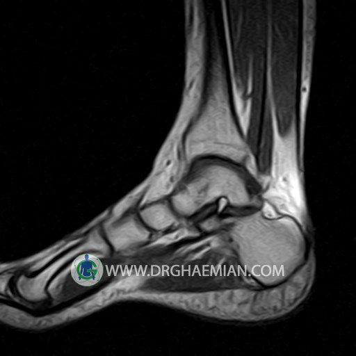
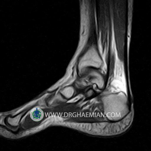
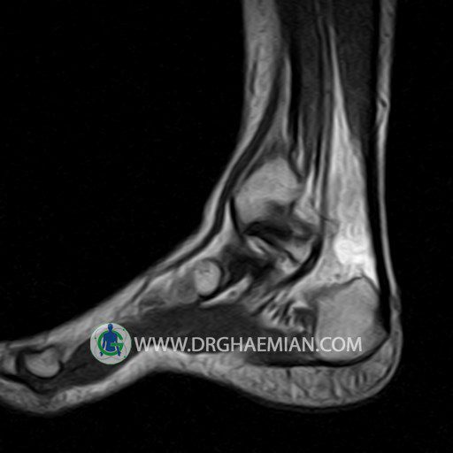
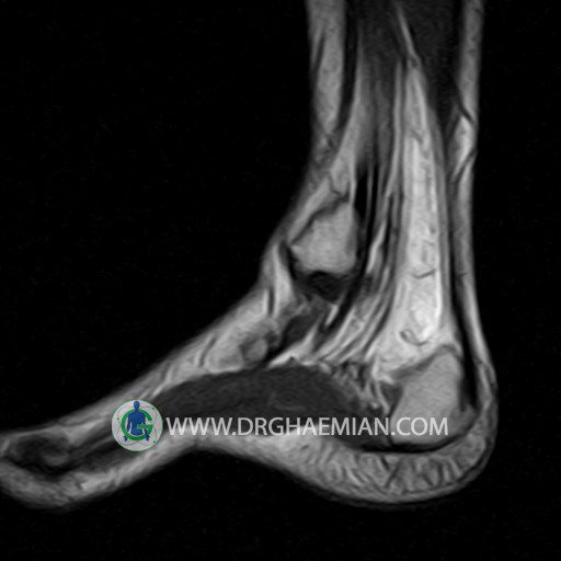
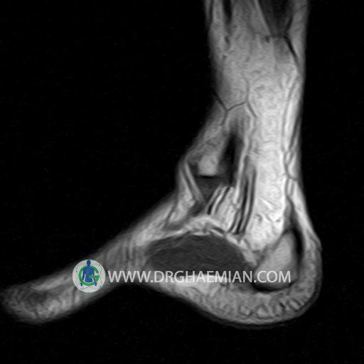
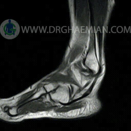
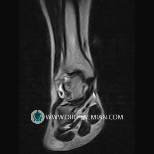
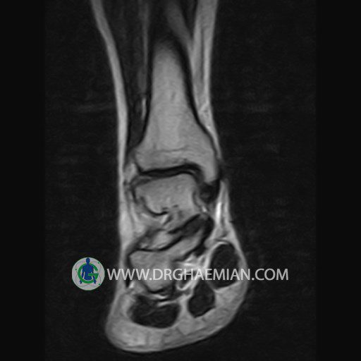
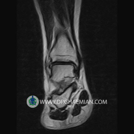
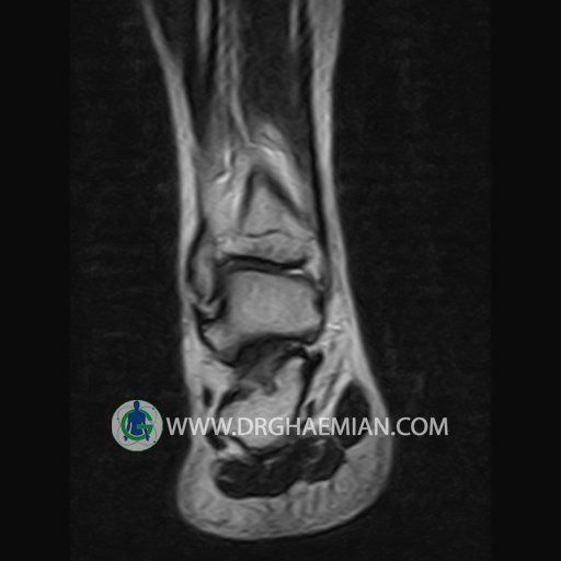
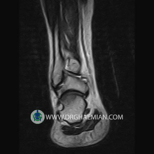
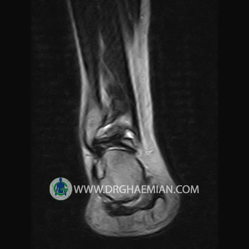
پزشکان اغلب از تصویربرداری ام آر آی برای تشخیص و درمان عارضه های پزشکی که فقط با استفاده از اشعه ایکس یا میدان مغناطیسی و امواج رادیویی قابل مشاهده است، استفاده می کنند. دستگاه ام آر آی تصاویر دقیق از ساختار های داخلی بدن ایجاد می کند. در این کیس تنوسینوویت مچ پا و تورم ملایم در اطراف مچ پا دیده می شود

گزارش پزشک :
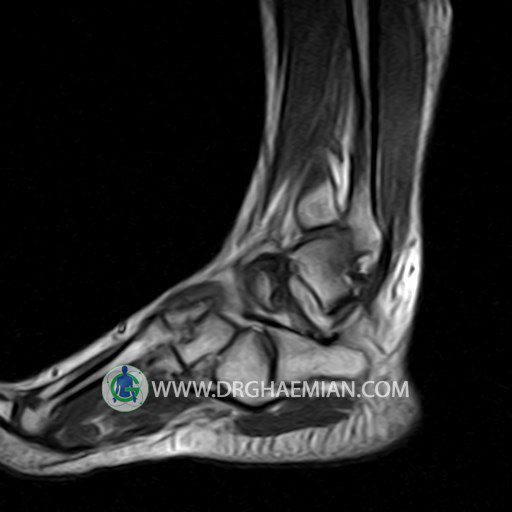
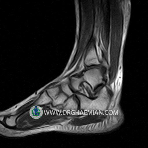
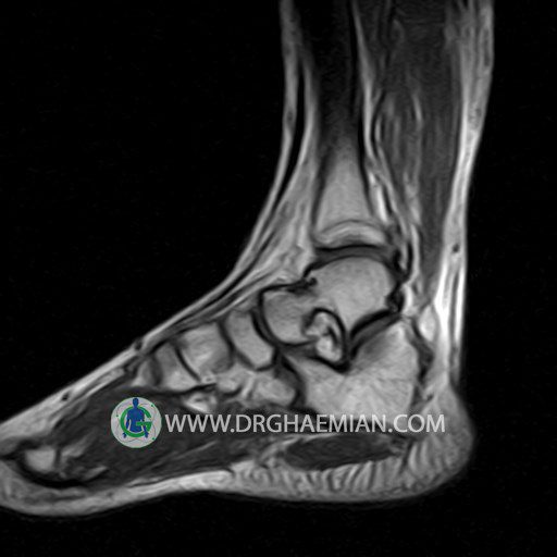
RIGHT ANKLE MRI
(Without contrast)
Technique :Axial T2 , coronal , sagittal T1 and T2 ,sagittal T2 fat sat .
REPORT:
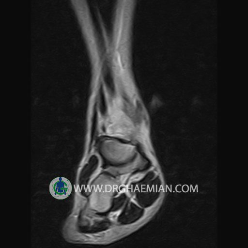
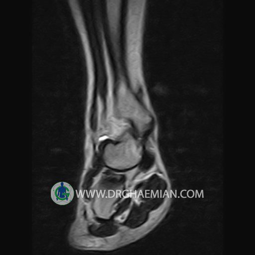
The bones comprising the ankle joint show normal position and configuration.
The bone marrow signal is normal.
The joint space is of normal width.
The cortex shows normal thickness and smooth contours , especially along the tibiotalar articular surfaces.
The lateral and medial ligaments are normal in their course , width and signal characteristics.
The talocalcaneal and talonavicular joints appear normal.
The achilles tendon is normal in its course , width and signal characteristics and the preachilles fat is clear.
Tarsal sinus is normal in shape and signal intensity.
– Mild soft tissue swelling around the lateral malleolus
– Fluid around the medial tendon suggestive for tenosynovitis
are seen
