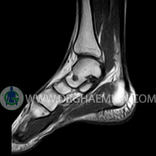
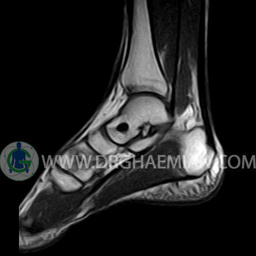
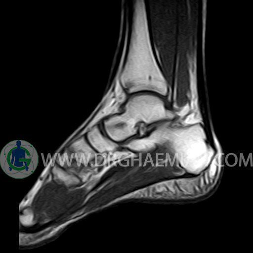
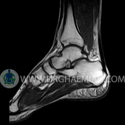
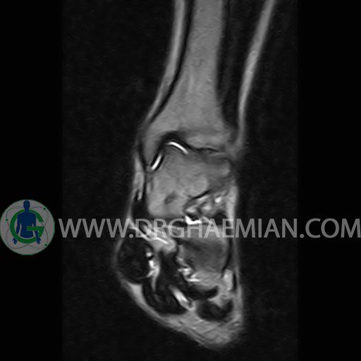
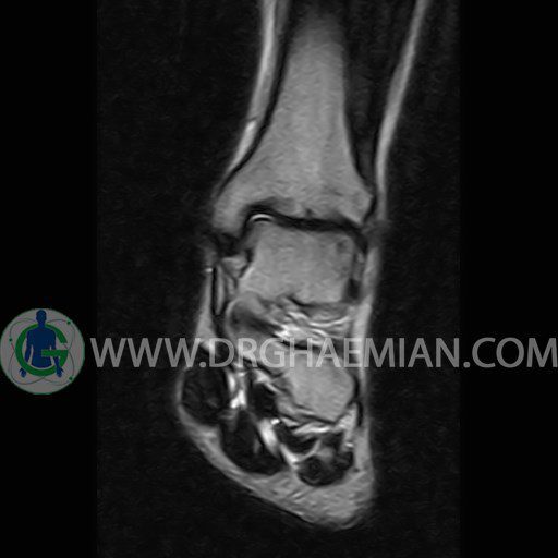
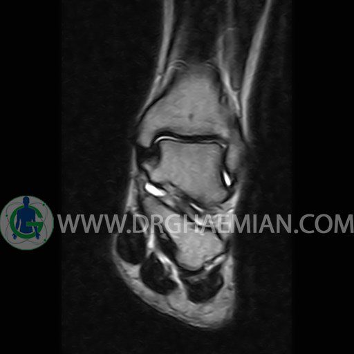
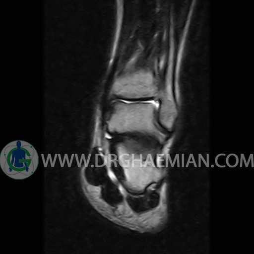
ام آر آی پا یک روش تصویربرداری از پا است که با استفاده از آهنرباهایی قوی تصاویری از پا ایجاد می کند. این تصاویر شامل قوزک، ساق پا و بافت های پیرامون آن ها می شود. ام آر آی از تشعشعات استفاده نمی کند. در این کیس گیرافتادگی خلفی مچ پا، سینوویت گذرا و کبودی استخوان دیده می شود.
گزارش پزشک :
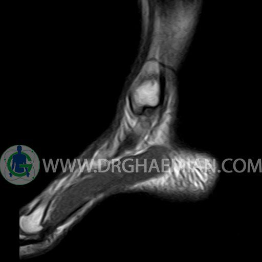
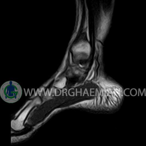
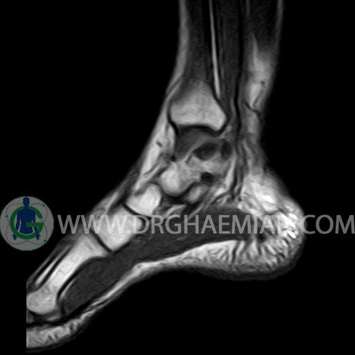
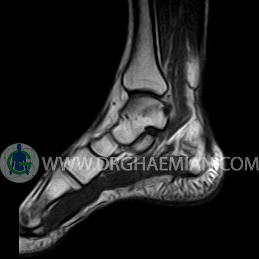
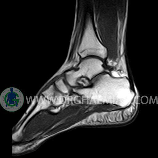
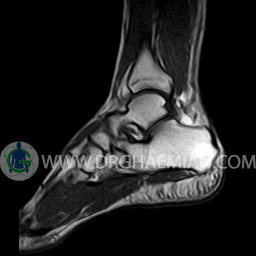
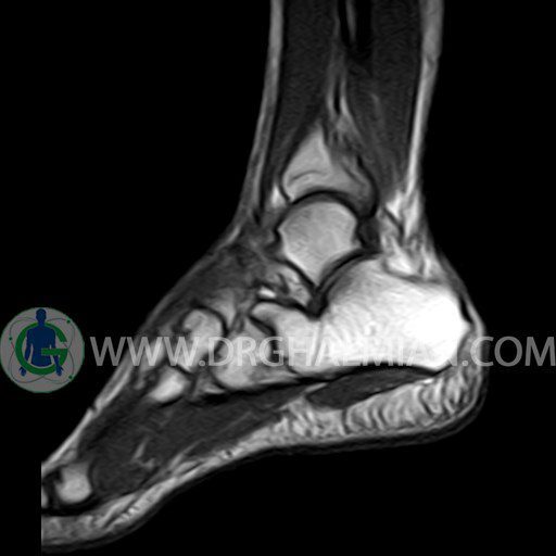
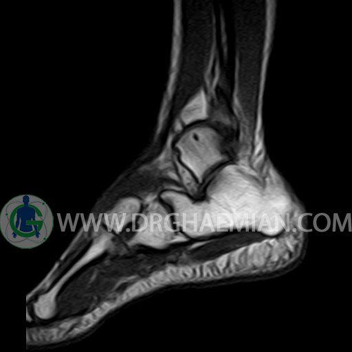
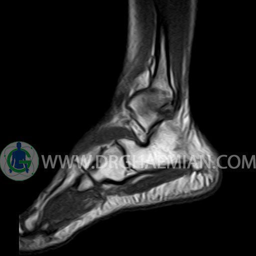
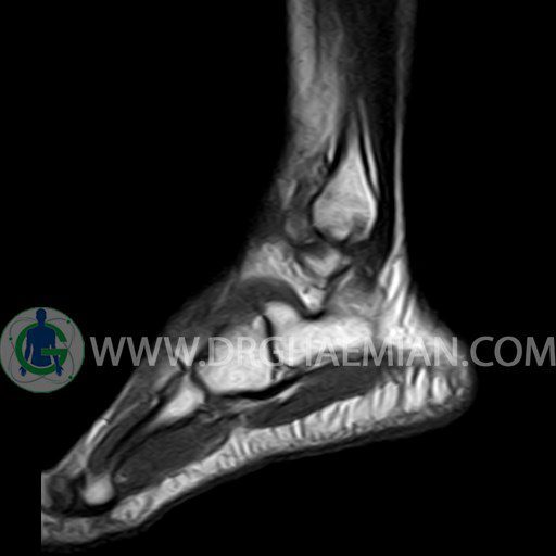
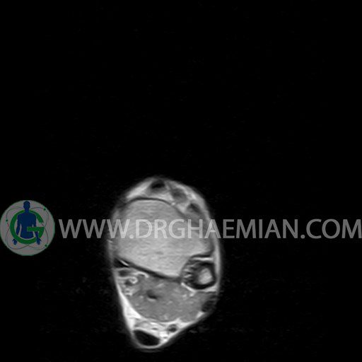
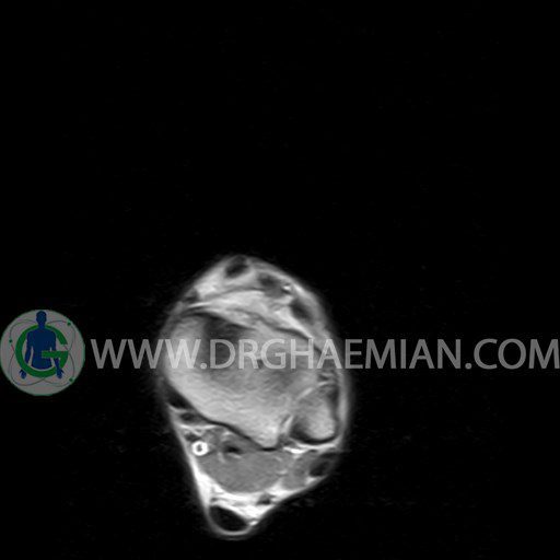
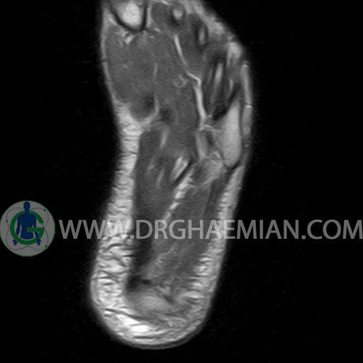
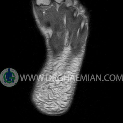
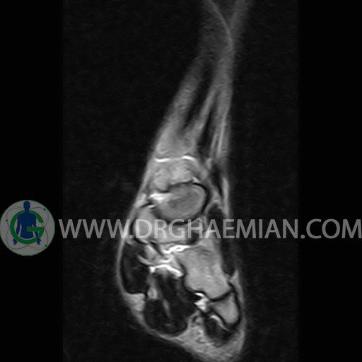
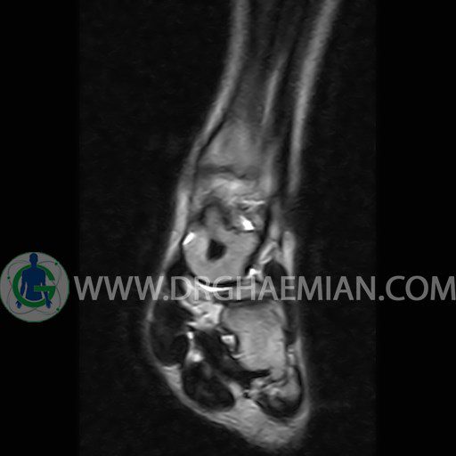
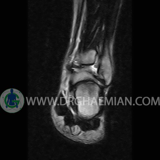
LEFT ANKLE MRI
(Without contrast)
Technique :Axial T2 , coronal , sagittal T1 and T2 ,sagittal T2 fat sat .
REPORT:
The bones comprising the ankle joint show normal position and configuration.
The joint space is of normal width.
The cortex shows normal thickness and smooth contours , especially along the tibiotalar articular surfaces.
The lateral and medial ligaments are normal in their course , width and signal characteristics.
The achilles tendon is normal in its course , width and signal characteristics and the preachilles fat is clear.
Tarsal sinus is normal in shape and signal intensity.
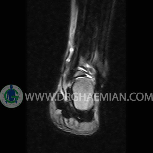
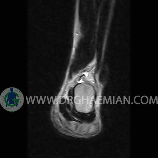
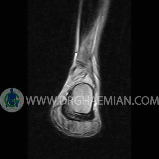
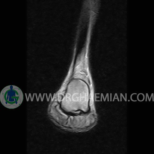
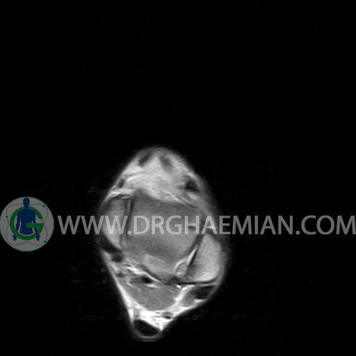
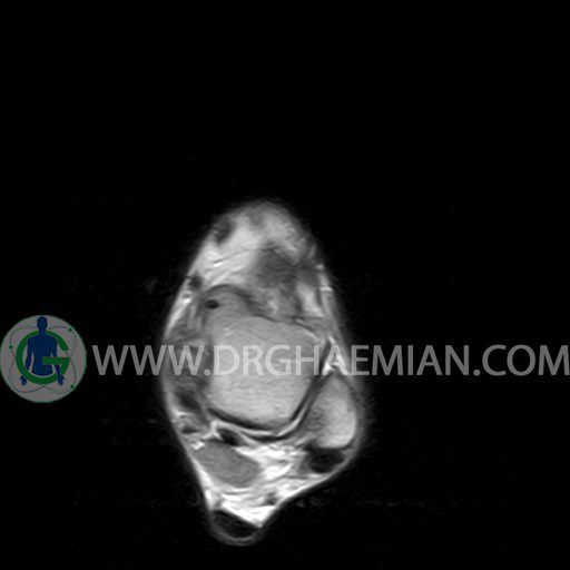
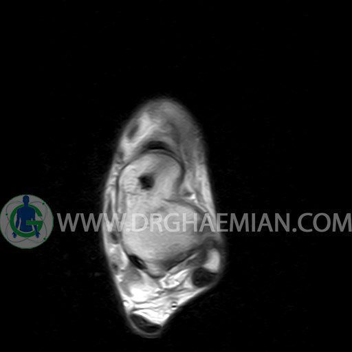
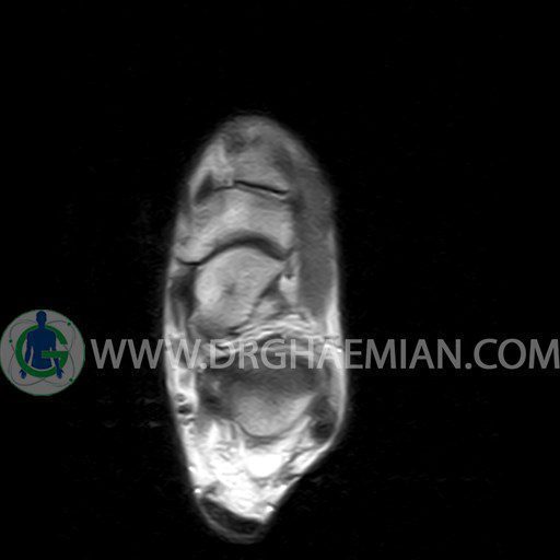
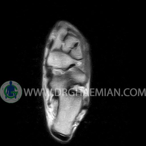
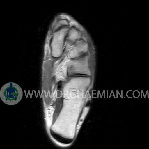
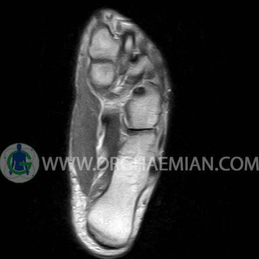
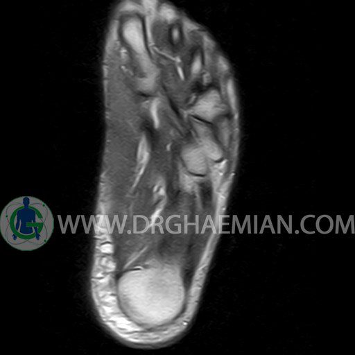
– Mild effusion in tibio – talo – calcaneus joint suggestive for transient synovitis
– Bone bruise in tibial insertion of talus
– Elongated lateral tubercle of talus ( stieda process ) with adjacent soft tissue swelling & edema in the retrocalcaneals bursa suggestive for posterior impingement
are seen
