ام آر آی مغز یک روش تصویربرداری است که با استفاده آهنربا های قوی و امواج رادیویی تصاویری از مغز و بافت های عصبی پیرامونی آن ایجاد می کند.
برخی از ام آر آی های مغز نیازمند رنگی مخصوص بنام کنتراست هستند. کنتراست معمولا از طریق رگی در دست یا بازو شما تزریق می شود و به رادیولوژیست کمک میکند تا برخی از قسمت های مغز را شفاف تر ببیند.
دلایل تجویز ام آر آی مغز
ام آر آی معز برای تشخیص یا نظارت خیلی از بیماری ها و عارضه هایی که بر روی مغز تاثیر دارند استفاده می شود. از جمله:
- نقص مادرزادی
- خونریزی (خونریزی ساب آراکنوئید یا خونریزی در خود بافت مغز)
- سابقه خانوادگی گرفتی رگ
- عفونت مثل آبسه مغزی
- تومورها (سرطانی یا غیر سرطانی)
- اختلالات های هورمونی (مثل آکرومگالی، گالاکتوره، سندرم کوشینگ)
- ام اس
- سکته مغزی
نوعی مخصوص از ام آر آی به نام ام آر ای (MRA) وجود دارد که تجویز می شود تا عروق خونی داخل مغز مشاهده شود.
REPORT :
The interhemispheric fissure is centered on the midline .
No abnormalities are seen in the basal ganglia , int . capsule, corpus callosum , Thalamus and tectal plate .
The sella and pituitary – pineal g .are normal and parasellar, suprasellar structures are unremarkable.
The cerebello pontine angle area appears normal on each side.
The internal acoustic meatus has normal width .
The orbital contents , falx , dura and calvaria are unremarkable .
Major intracranial vascular structures , pericavernous spaces and visualized intracranial nerve complex are normal .
The paranasal sinuses are clear and aerated with no evidence of bone erosion or destruction, fluid collection , cystic retention and mucosal thickening.
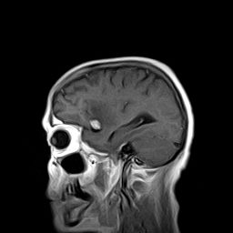
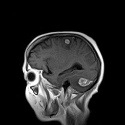
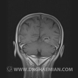
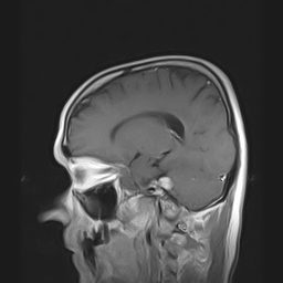
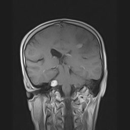
– Multiple , intra – axial mass lesion in left frontal lobe ( 11 mm ), right temporal
pole( 14 mm ) & in left cerebellum ( 28 mm ) , with peripehral vasogenic edema
& with post contrast bright enhancement suggestive for brain metastasis ( without significant change to previous MRI )
REPORT :
The interhemispheric fissure is centered on the midline .
No abnormalities are seen in the basal ganglia , int . capsule, corpus callosum , Thalamus and tectal plate .
The brain stem , cerebellum show no abnormal changes in signal characteristics.
The sella and pituitary – pineal g .are normal and parasellar, suprasellar structures are unremarkable.
The cerebello pontine angle area appears normal on each side.
The internal acoustic meatus has normal width .
The orbital contents , falx , dura and calvaria are unremarkable .
Major intracranial vascular structures , pericavernous spaces and visualized intracranial nerve complex are normal .
The paranasal sinuses are clear and aerated with no evidence of bone erosion or destruction, fluid collection , cystic retention and mucosal thickening.
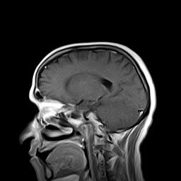
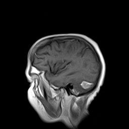
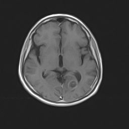
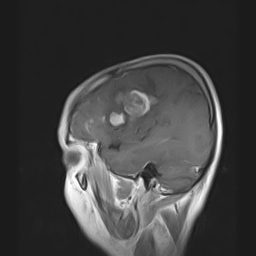
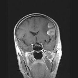
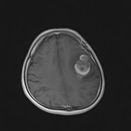
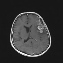
– A well – defined solid cystic mass lesion in left occipital lobe , with peripheral vasogenic edema & with post contrast heterogeneous ring enhancement & a second small mass lesion in right occipital lobe with post contrast enhancement suggestive for cerebral metastasis

REPORT :
The interhemispheric fissure is centered on the midline .
No abnormalities are seen in the basal ganglia , int . capsule, corpus callosum , Thalamus and tectal plate .
The sella and pituitary – pineal g .are normal and parasellar, suprasellar structures are unremarkable.
The internal acoustic meatus has normal width .
The orbital contents , falx , dura and calvaria are unremarkable .
Major intracranial vascular structures , pericavernous spaces and visualized intracranial nerve complex are normal .
The paranasal sinuses are clear and aerated with no evidence of bone erosion or destruction, fluid collection , cystic retention and mucosal thickening.
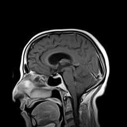
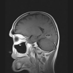
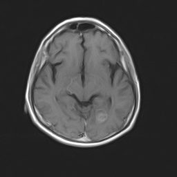
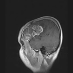
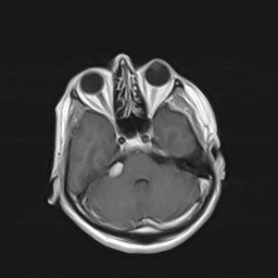
– Left parietofrontal craniotomy
– An ill defined – intra axial mass lesion in left parietofrontal lobes with post contrast heterogeneous ring enhancement , with peripheral vasogenic edema & with mass effect on left lateral ventricle suggestive for recurrent/ remnant tumor
– A well defined – extra axial mass lesion in right CPA with post contrast bright enhancement suggestive for acoustic neuroma
