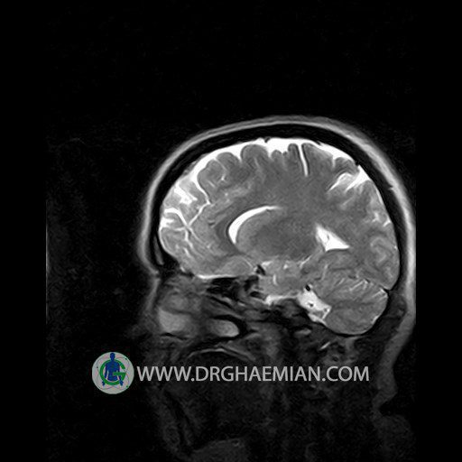
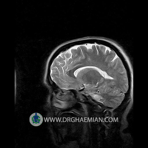
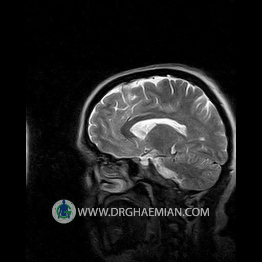
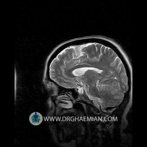
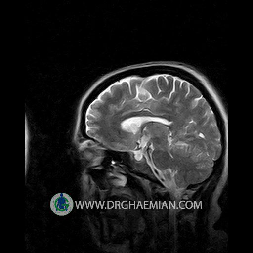
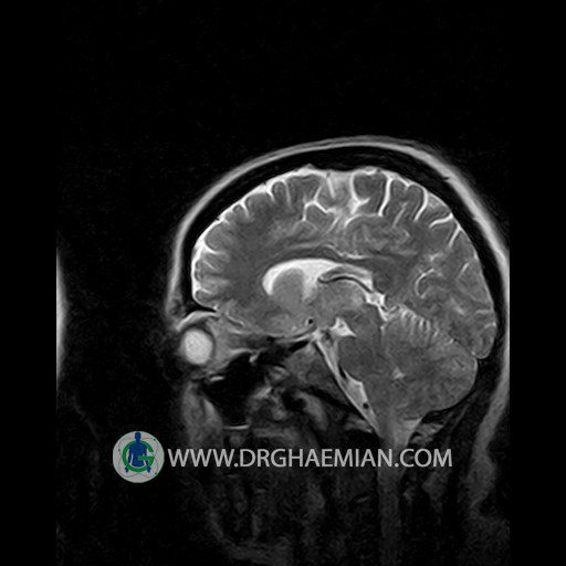
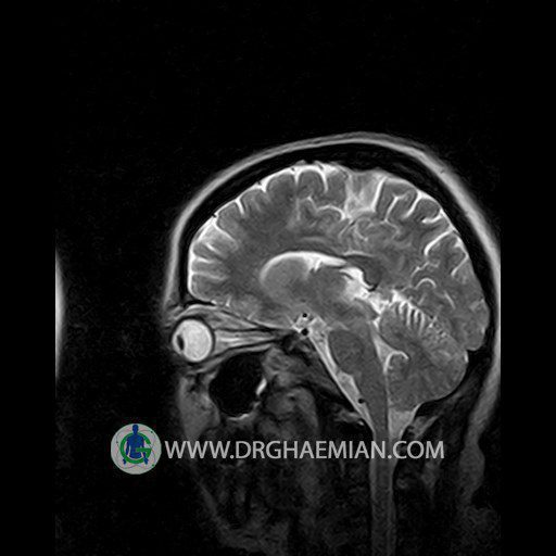
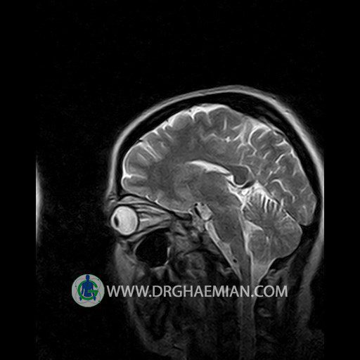
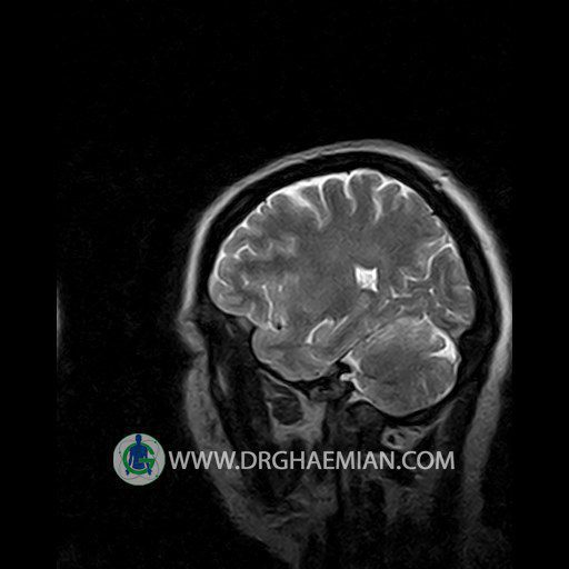
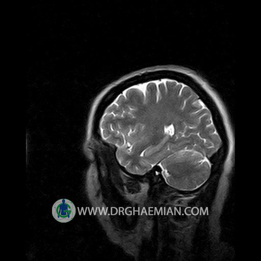
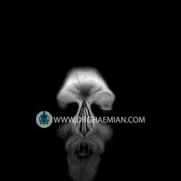
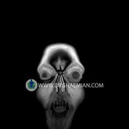
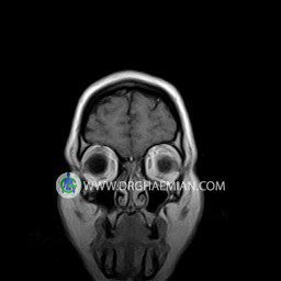
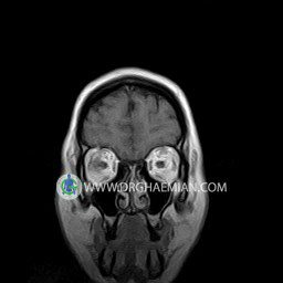
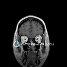
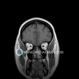
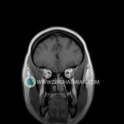
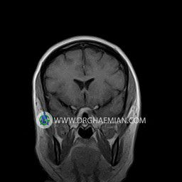
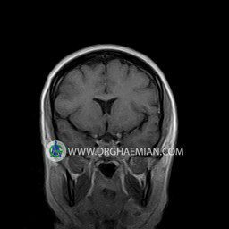
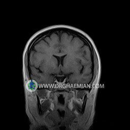
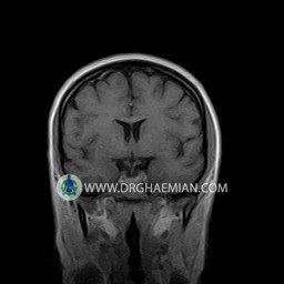
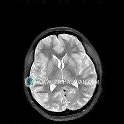
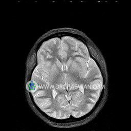
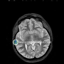
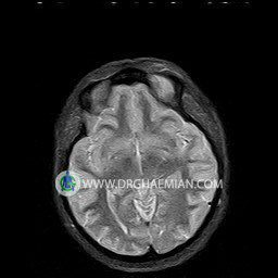
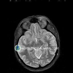
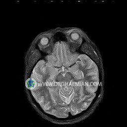
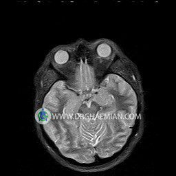
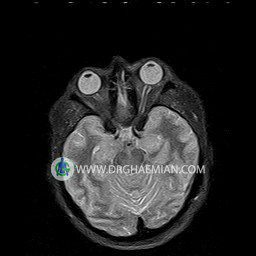
پزشکان اغلب از تصویربرداری ام آر آی برای تشخیص و درمان عارضه های پزشکی که فقط با استفاده از اشعه ایکس یا میدان مغناطیسی و امواج رادیویی قابل مشاهده است، استفاده می کنند. دستگاه ام آر آی تصاویر دقیق از ساختار های داخلی بدن ایجاد می کند. در این کیس نوریت اوربیت چب و سلای خالی بیمار مشاهده می شود.
گزارش پزشک :
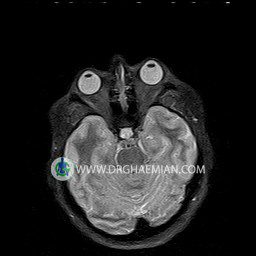
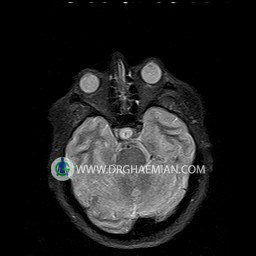
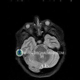
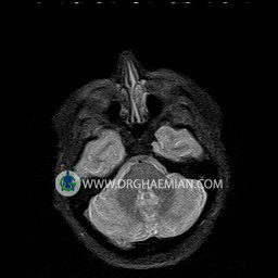
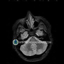
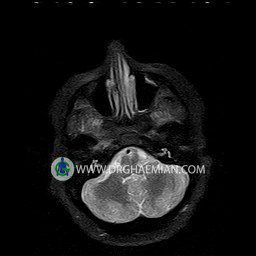
ORBIT MRI
(with and without contrast)
Technique:Axial T1 , Axial , sagittal , coronal FSE T2 , coronal T1, sagittal fat sat T2 , Axial , sagittal T1 post Gd .
REPORT :
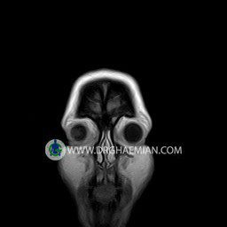
The both orbit are symmetrical and of normal size , with normal development of the orbital cones .
The bony orbital walls show a normal configuration with smooth and, sharp margins .
No foci of bone destruction , no circumscribed expansion of the bony or soft – tissue components of the orbital are evident .
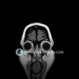
The globes are symmetrical and of normal size and the ocular contents show normal signal characteristics .
The ocular walls are smooth , sharply defined , and of normal thickness .
The retrobulbar fat, ophthalmic vein and lacrimal apparatus are unremarkable .
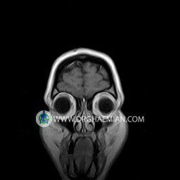
Evaluable portions of the neurocranium and paranasal sinuses show no abnormalities .
No seen any evidence of ocular herniation
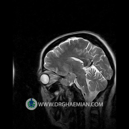
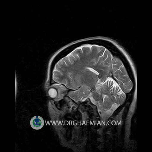
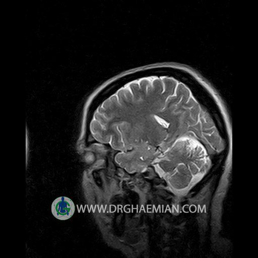
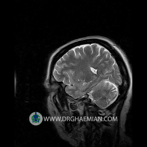
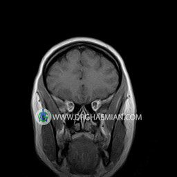
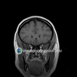
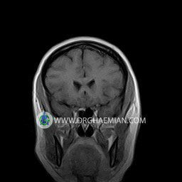
– Small fluid around the left optic nerve with mild edema suggestive for left optic neuritis
– Extension of suprasella cistern to sella with thin pituitary gland in floor of sella ( empty sella )
are seen
REPORTED BY :Dr DrNaser. Ghaemian.
