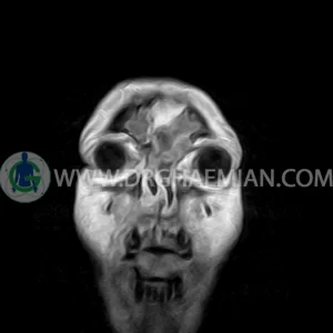
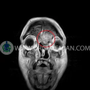
ام آر آی صورت و سینوس با استفاده از آهرنباها و امواج رادیویی تصاویری از اوربیت ها، ساختاری های صورت، عضلات و بافت های اطراف آن ایجاد می کند. در این کیس توده سینوس جلویی، سلای نسبتا خالی و ضخیم شدن مخاطی دیده می شود.
گزارش پزشک
CERVICAL & SINUS SPINE MRI
(with & without contrast)
Technique : Sagittal T1 , T2 , Axial T2 .
REPORT:
The cervical spine shows a smooth lordotic curvature with normal alignment.
The intervertebral disk spaces are normal height and signal intensity .
The bony spinal canal has normal width which , neural foramina and posterior element displays normal nature .
The visualized spinal cord is normal .
Predense distance is normal .
Paravertebral stripe is normal in shape and signal intensity .
The nasal turbinates show a normal arrangement and normal signal intensity.
The pharynx and imaged parapharyngeal structures petrygoid lamina and fossa show no abnormalities.
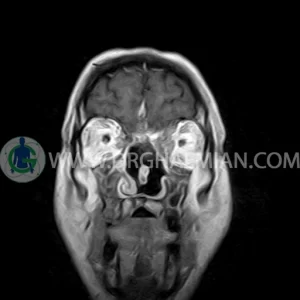
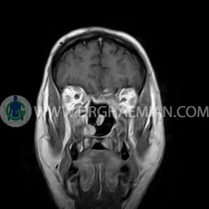
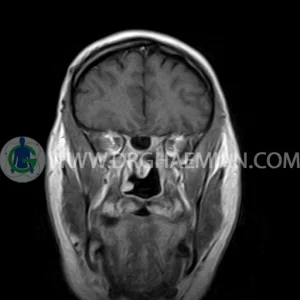
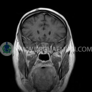
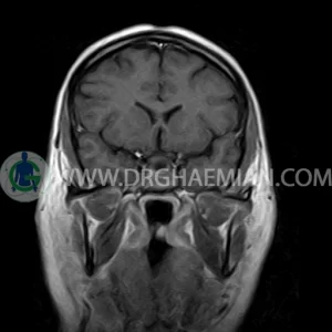
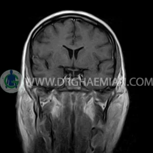
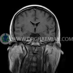
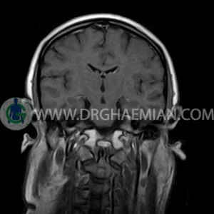
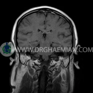
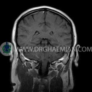
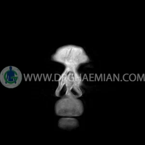
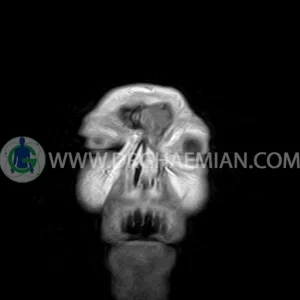
– Post op. changes in nasal cavity & in left ethmoid & maxillary sinuses
– An ill – defined lobulated solid cystic mass lesion ( 30 x 45 mm ) in left frontal sinus with extention to left orbit , let proptosis , destruction of left frontal & medial wall of left orbit & with post contrast heterogeneous enhancement suggestive for recurrent / remnant tumor
– Extension of suprasella cistern to sella with thin pituitary gland in floor of sella ( partial empty sella )
– Mucosal thickening with fluid collection in sphenoid , right frontal & right maxillary sinuses
are seen
