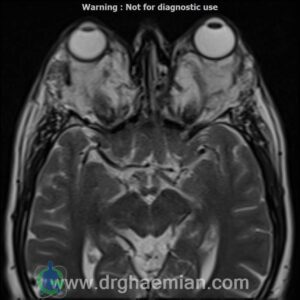
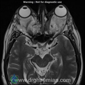
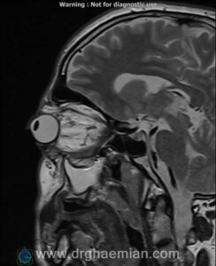
ام آر آی اوربیت با استفاده از آهنربا و امواج رادیویی تصاویری از اوربیت ها، اعصاب، عضلات و بافت های اطراف آن ایجاد می کند. در برخی از موارد ماده حاجب یا کنتراستی را می توان از طریق سرم تزریق کرد تا به نمایش بهتر برخی از ساختارها یا موارد غیرعادی کمک شود.در این کیس اُفتالموپاتی تیروئیدی چشم…. دیده میشود

گزارش پزشک
MRI OF ORBIT (WITHOUT IV GADOLINIUM)
1.5 Tesla MR System
Multislice, multiplanar and multisequence MR images reveal:
Lacrimal glands: unremarkable
Globes: normal
Optic nerve sheath complexes: normal
Optic chiasma and tracts: normal
Extra-ocular muscles: Bilateral enlargement of the medial and inferior rectus muscles with tendon sparing.
Intraconal and extraconal retro-orbital fat: Enlargement of intraorbital fat without abnormal signal intensity.
Superior orbital fissures: normal
Optic canals: normal
Bones and marrow: Evidence of prior surgery involving the lateral wall of the right orbit.
Visualized neurocranium: unremarkable
Paranasal sinuses: Mucosal thickening in both maxillary sinuses.
Additional finding: Significant bilateral proptosis.
Impression
Significant bilateral proptosis with bilateral enlargement of the medial and inferior rectus muscles with tendon sparing, along with increased intraorbital fat—findings highly suggestive of thyroid ophthalmopathy.
Post-surgical changes involving the lateral wall of the right orbit.
Mucosal thickening in both maxillary sinuses.
Otherwise unremarkable orbital MRI without contrast.