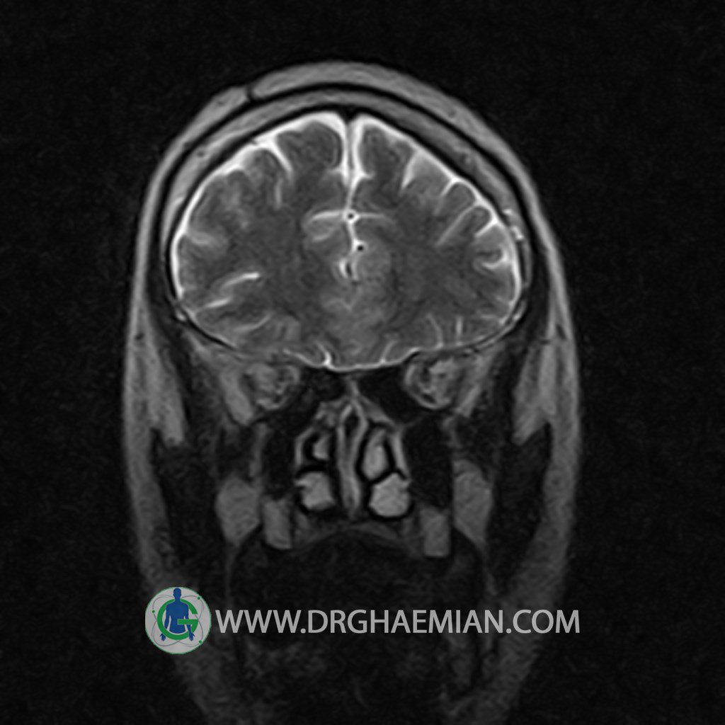
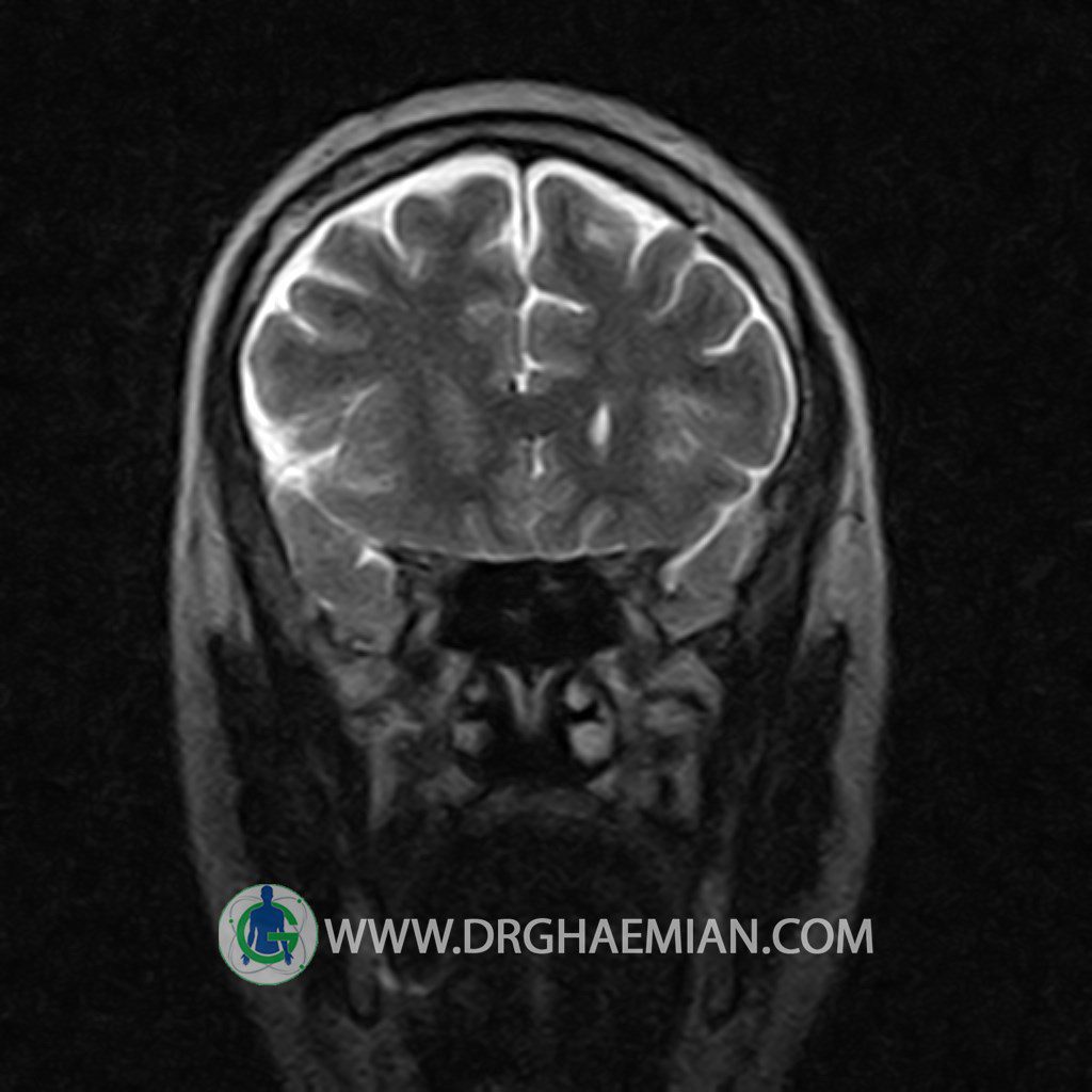
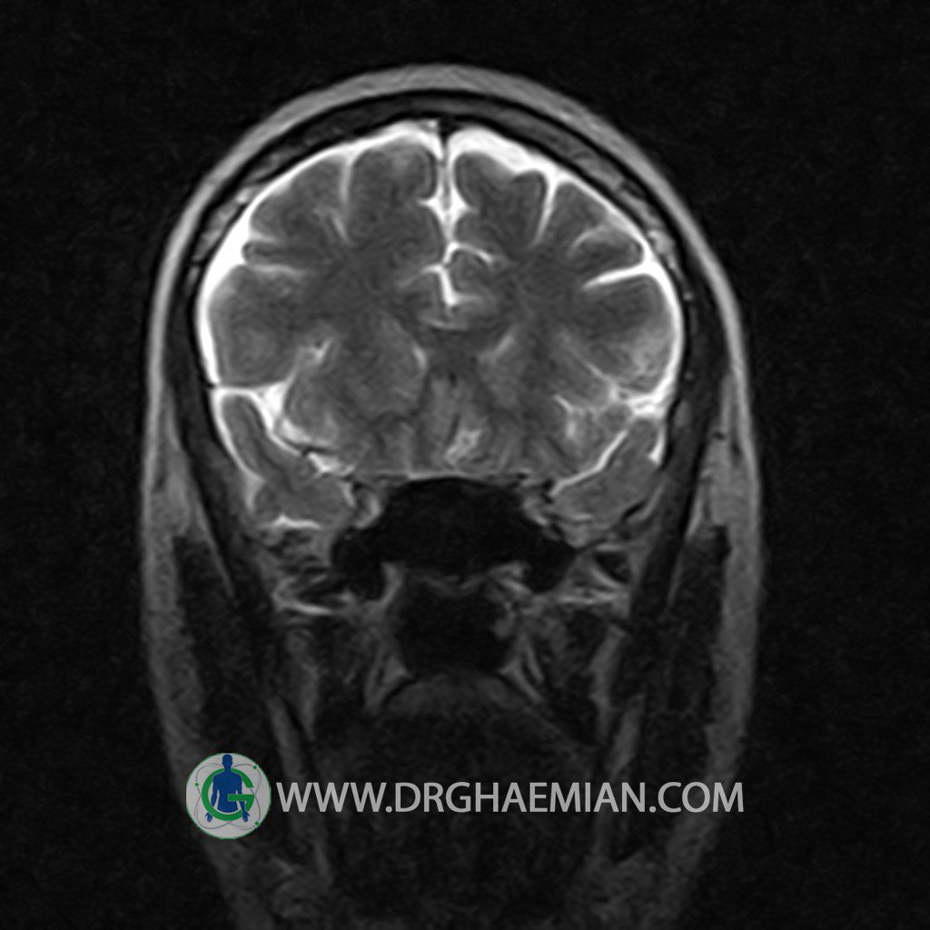
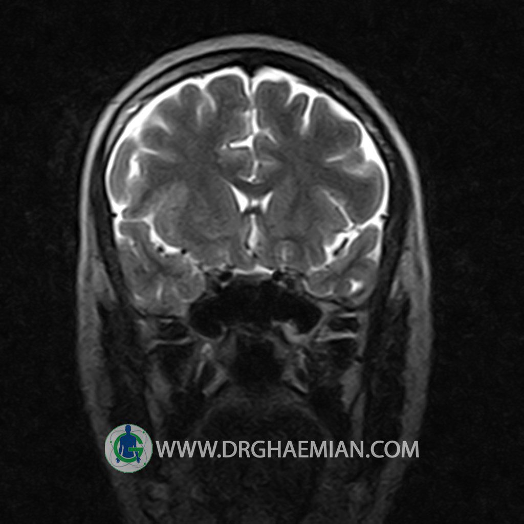
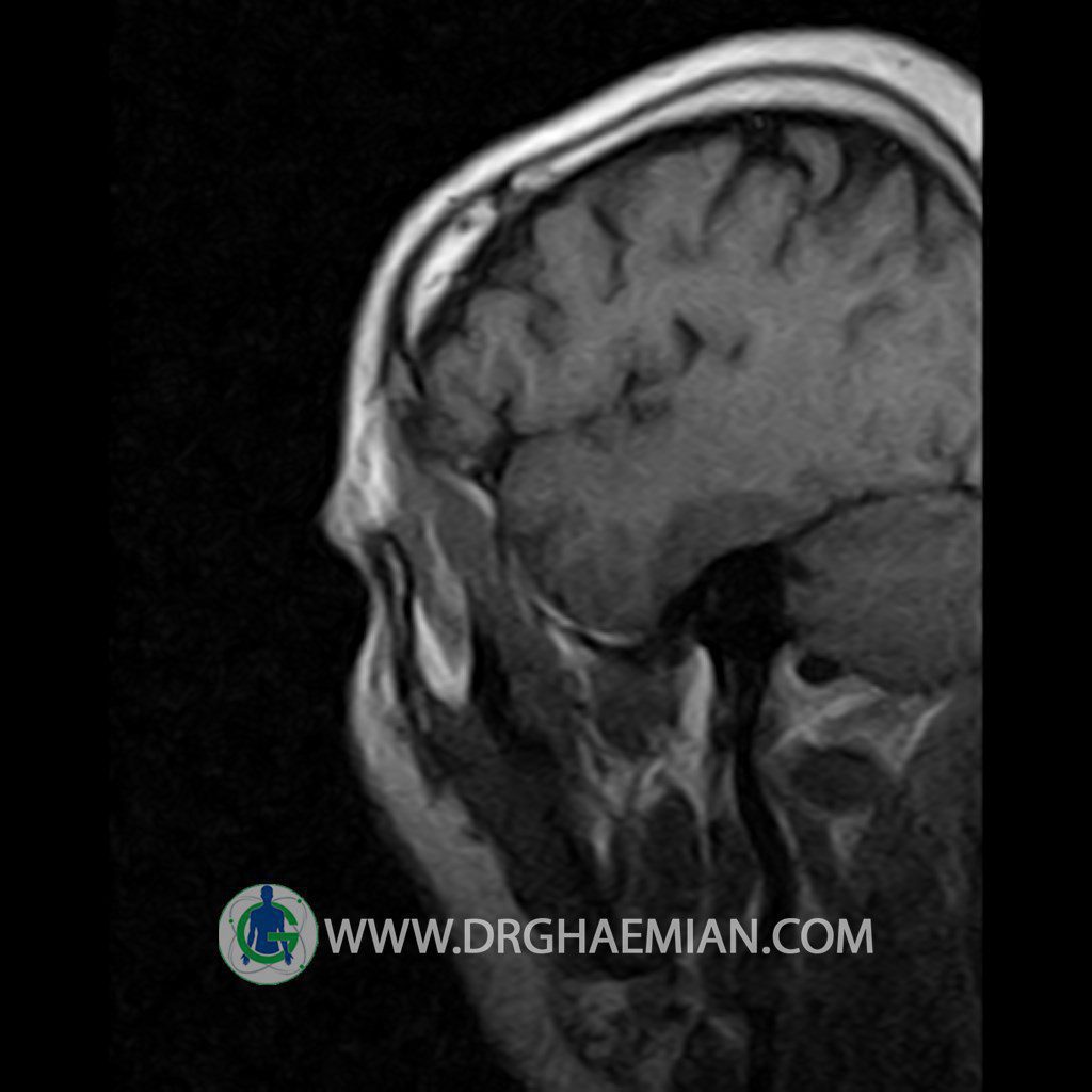
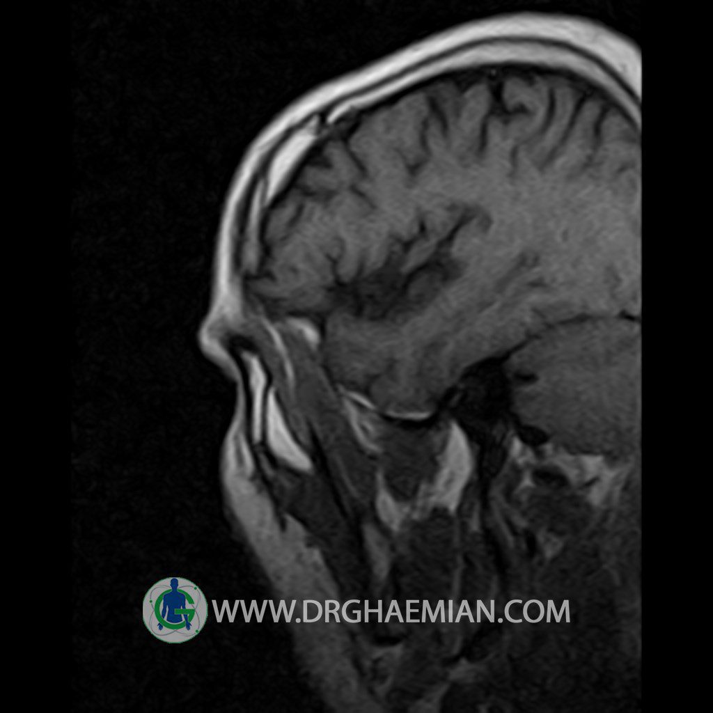
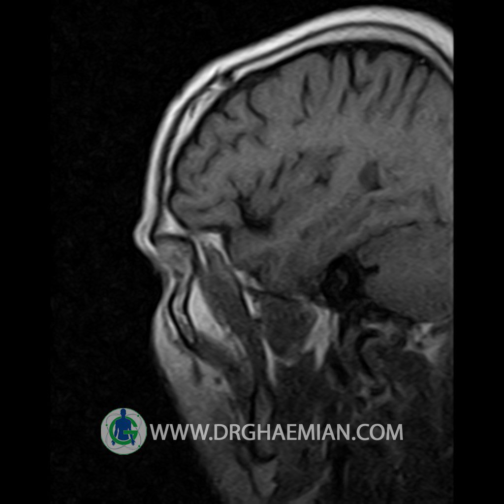
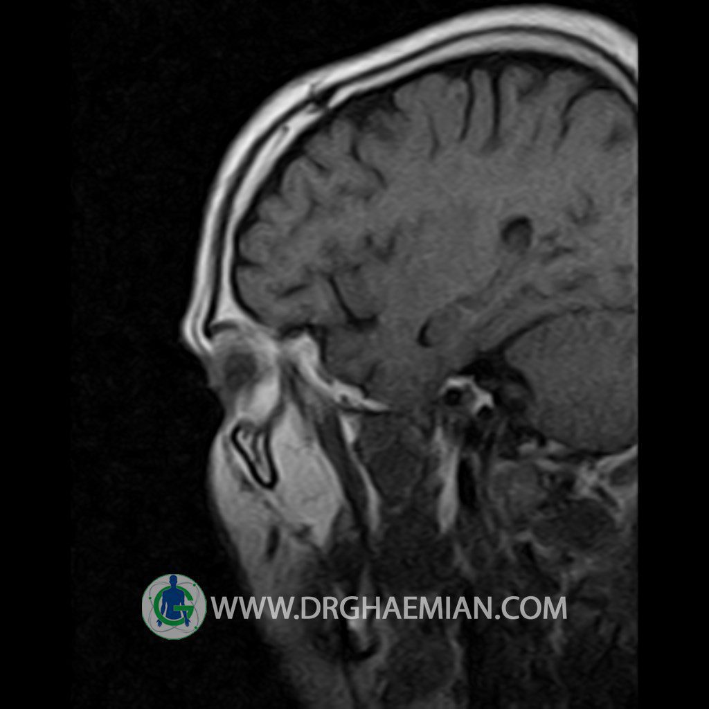
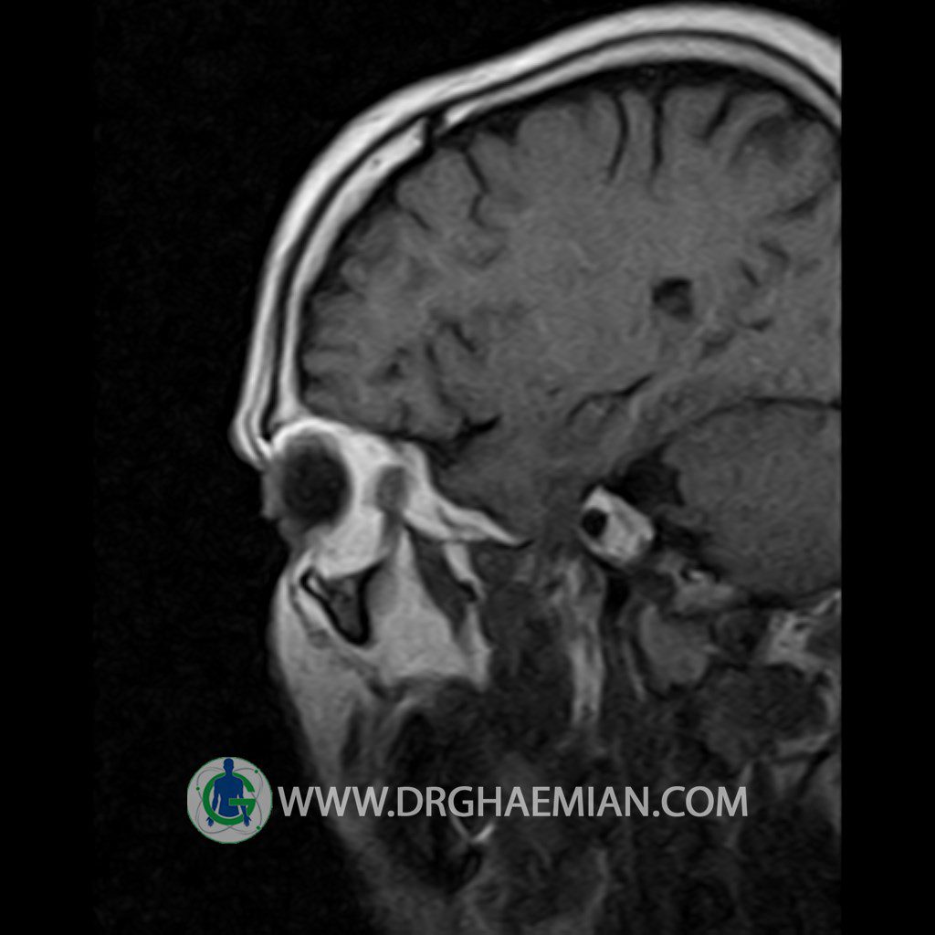
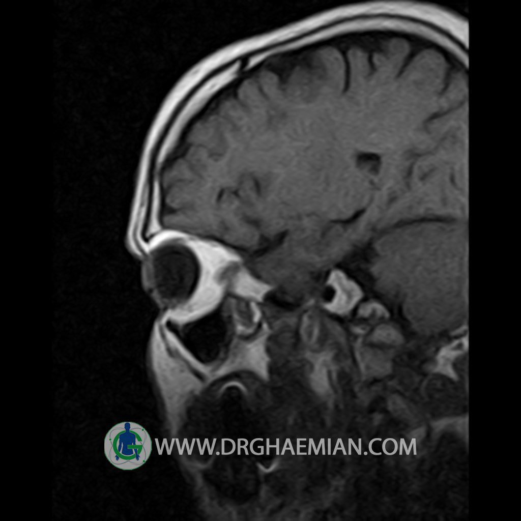
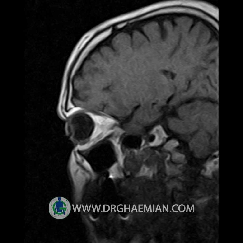
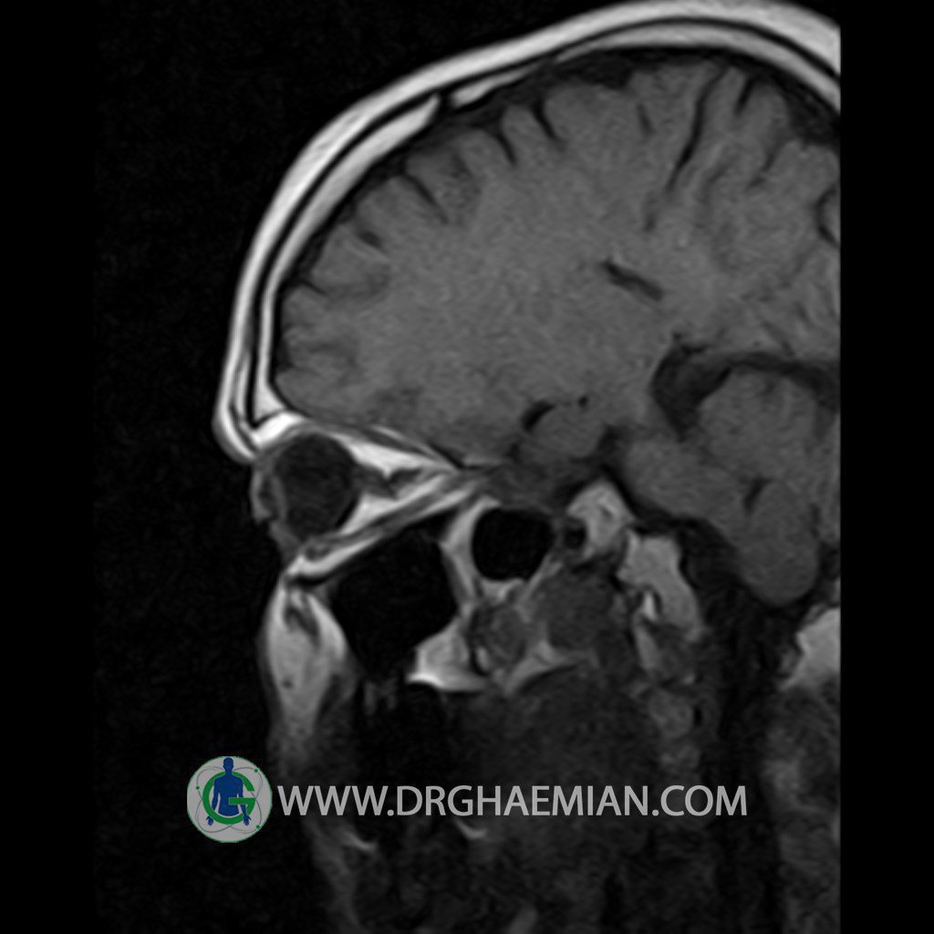
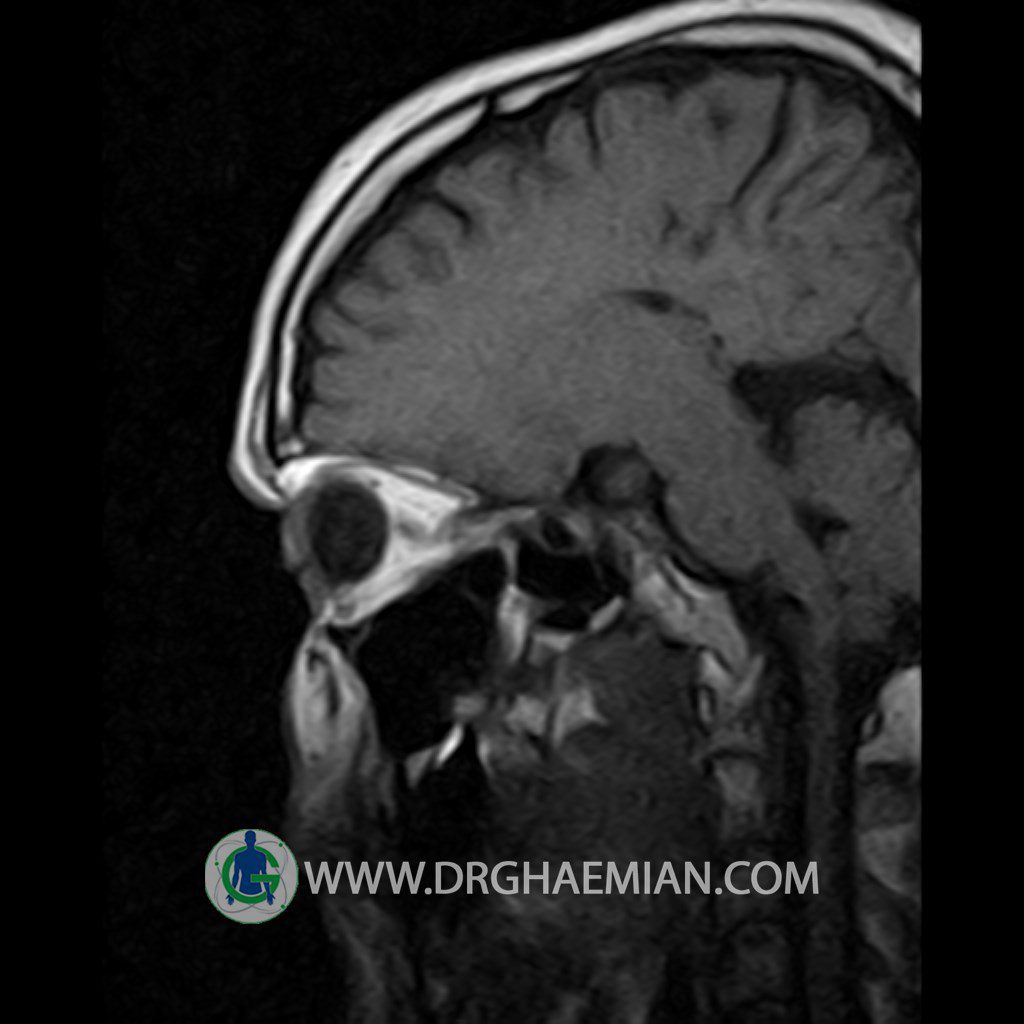
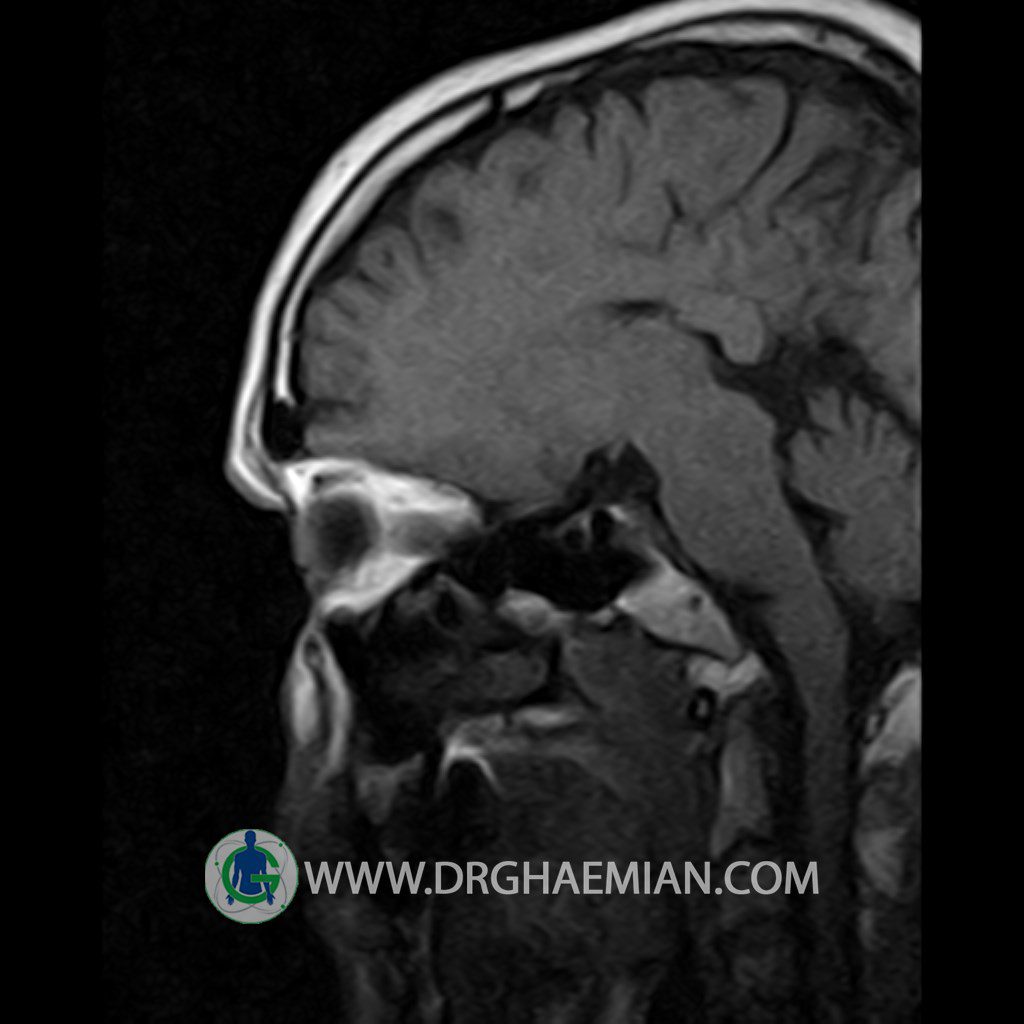
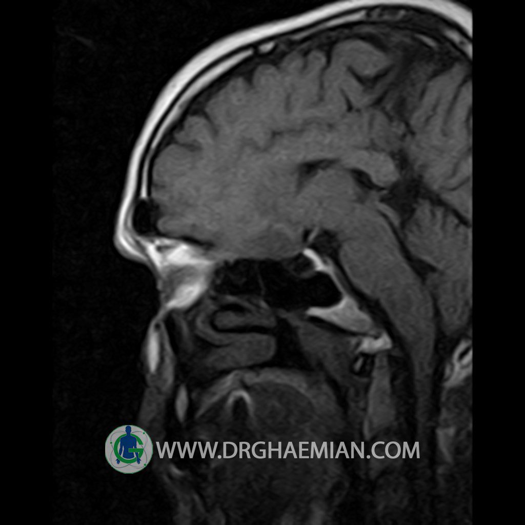
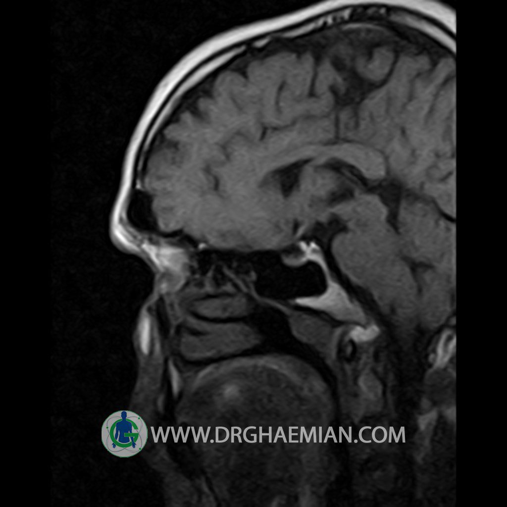
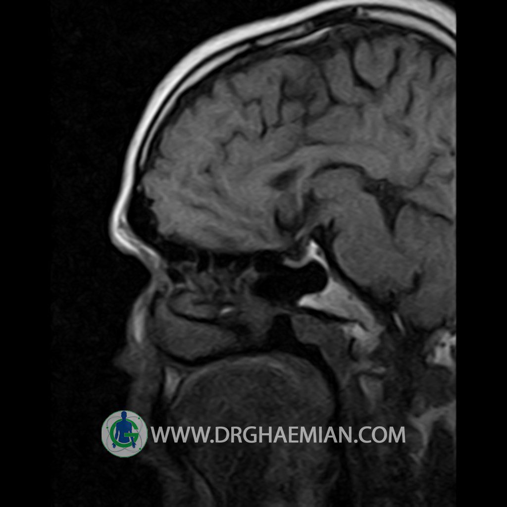
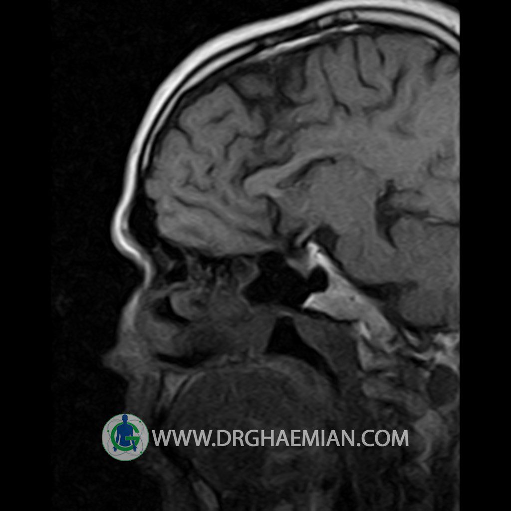
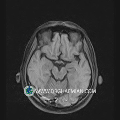
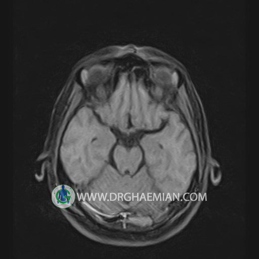
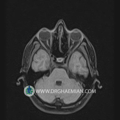
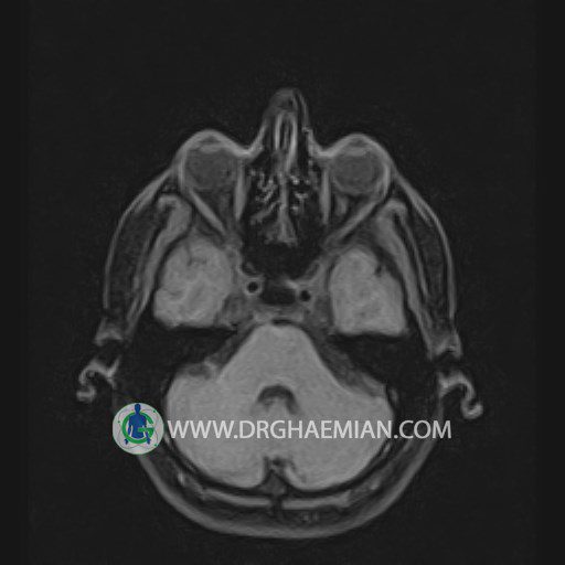
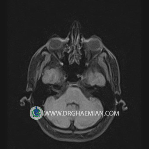
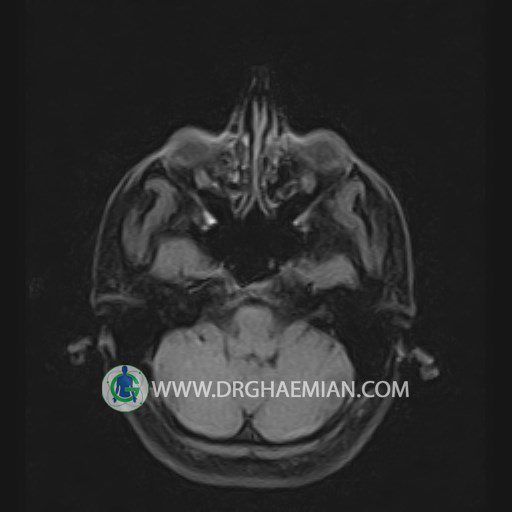
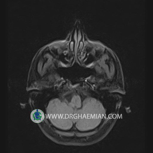
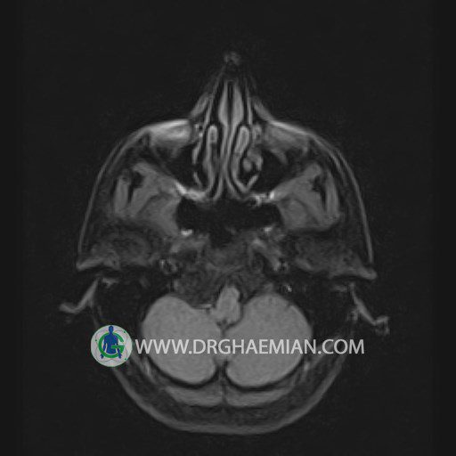
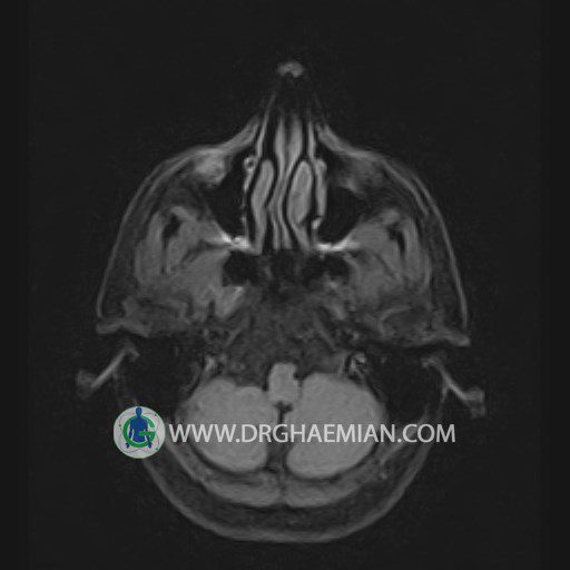
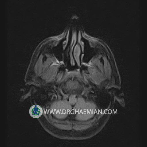
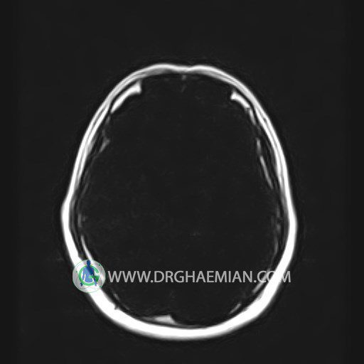
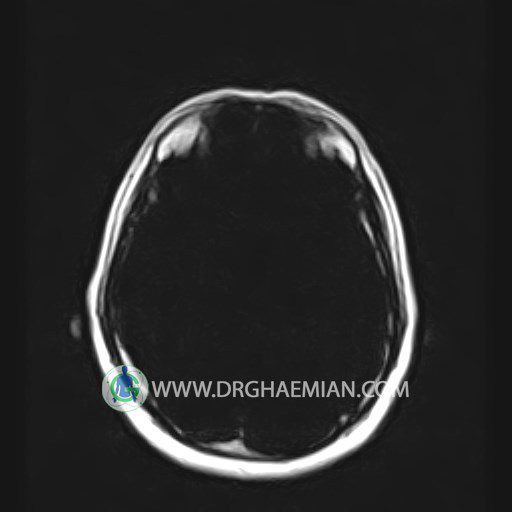
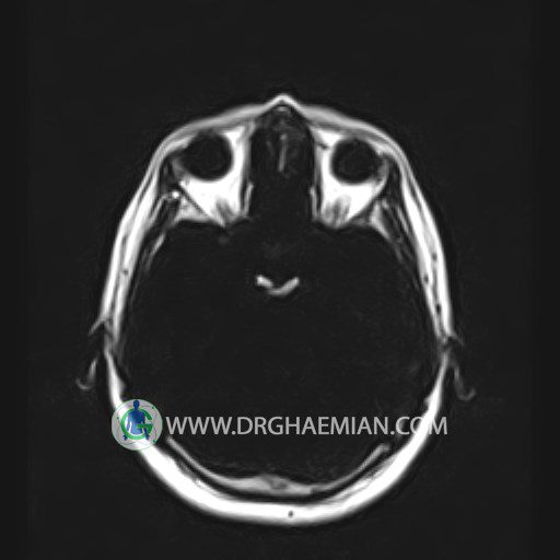
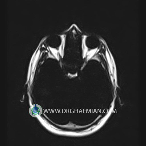
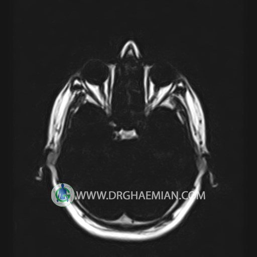
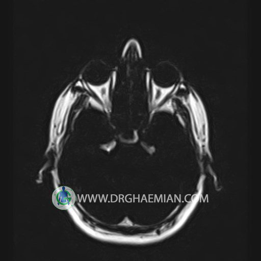
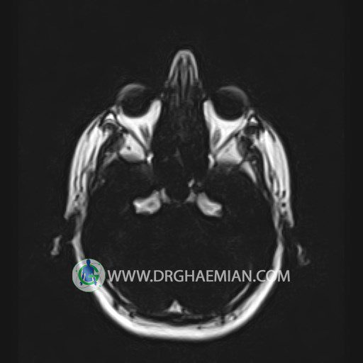
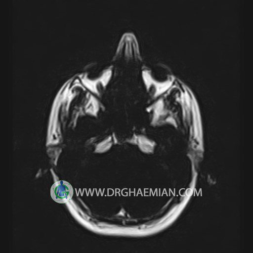
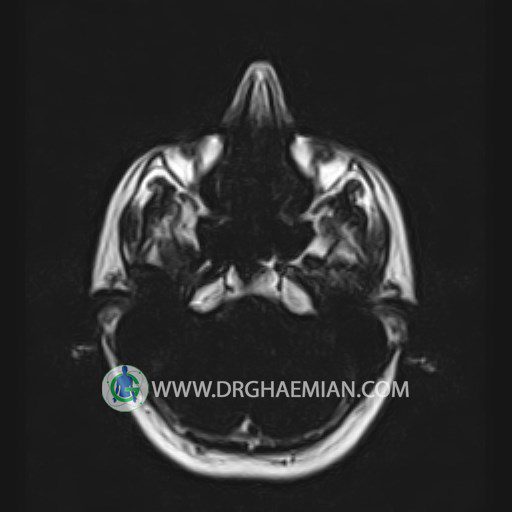
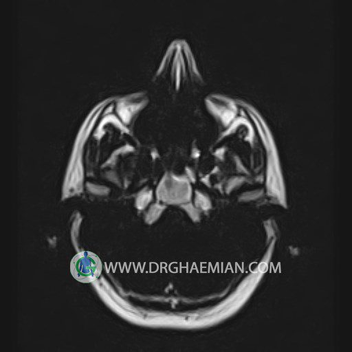
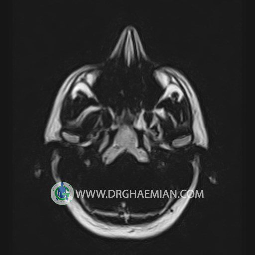
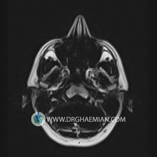
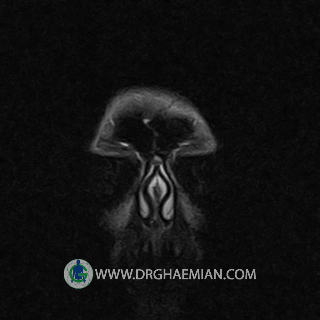
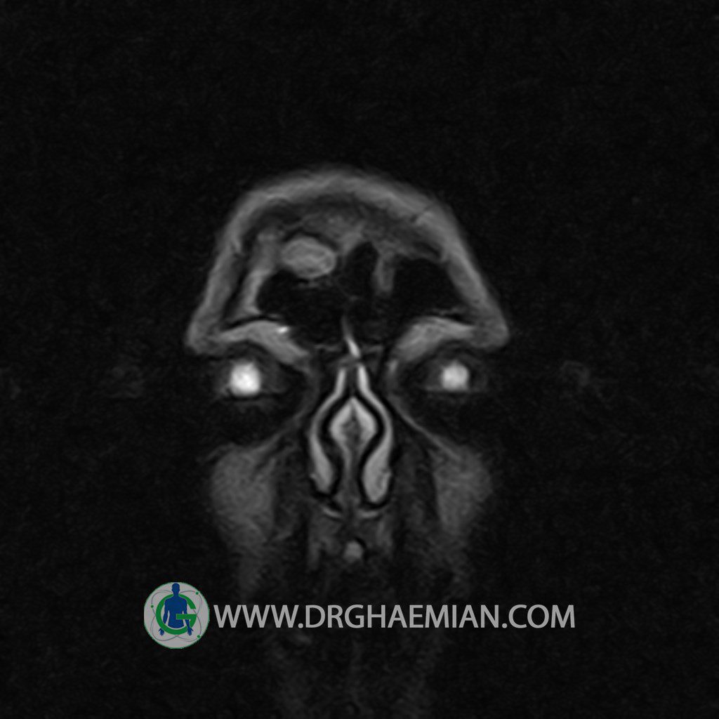
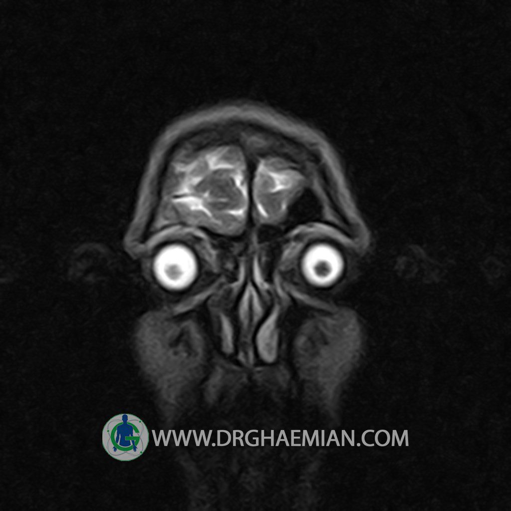
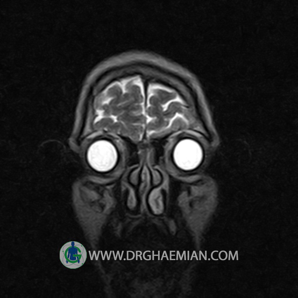
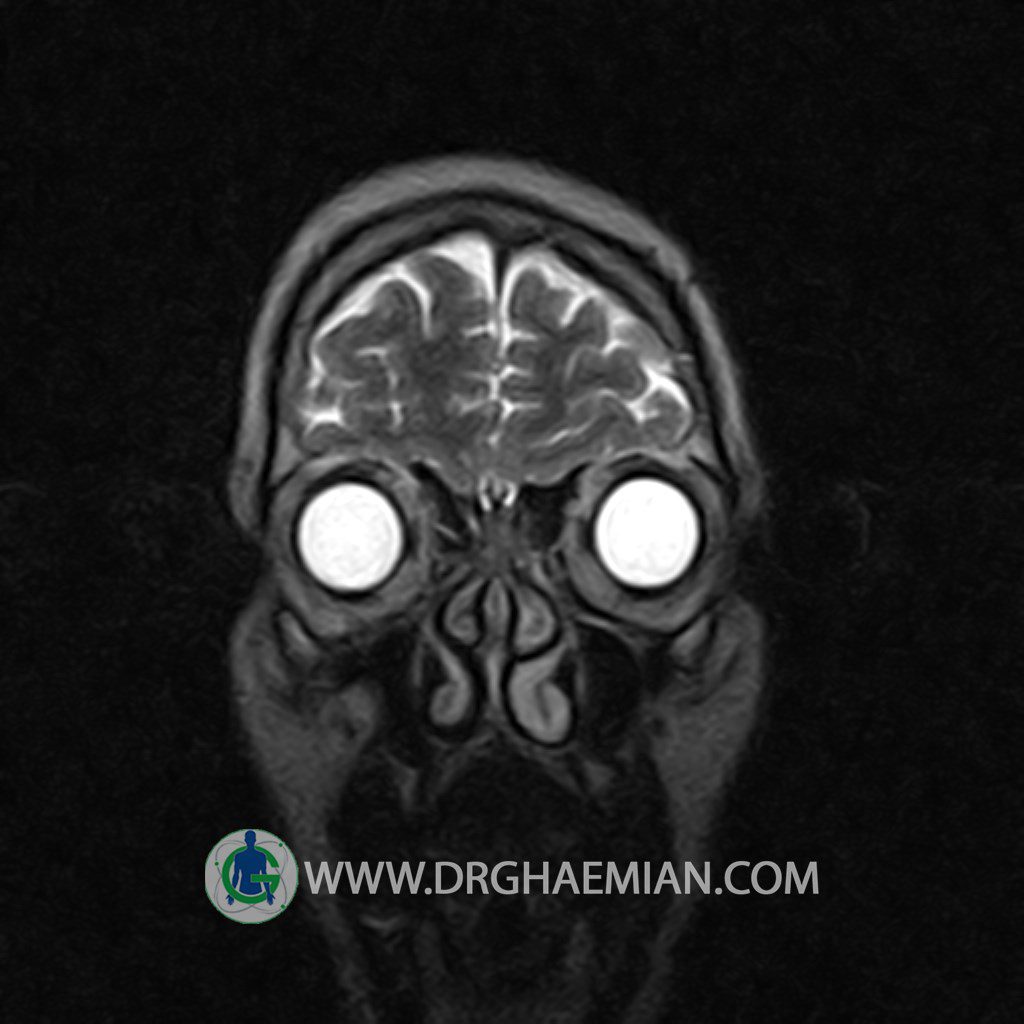
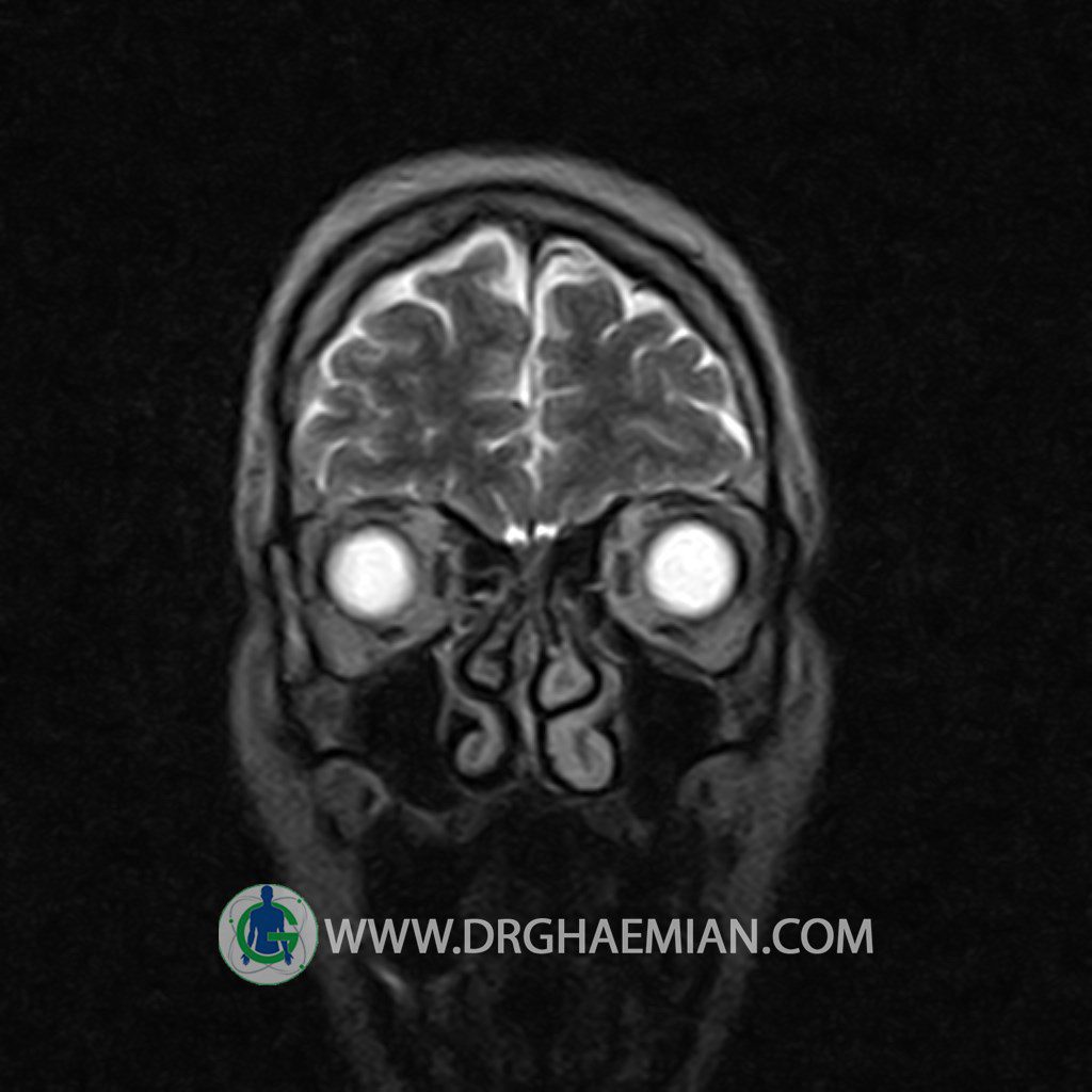
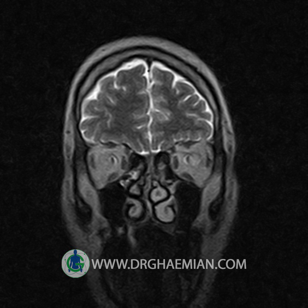
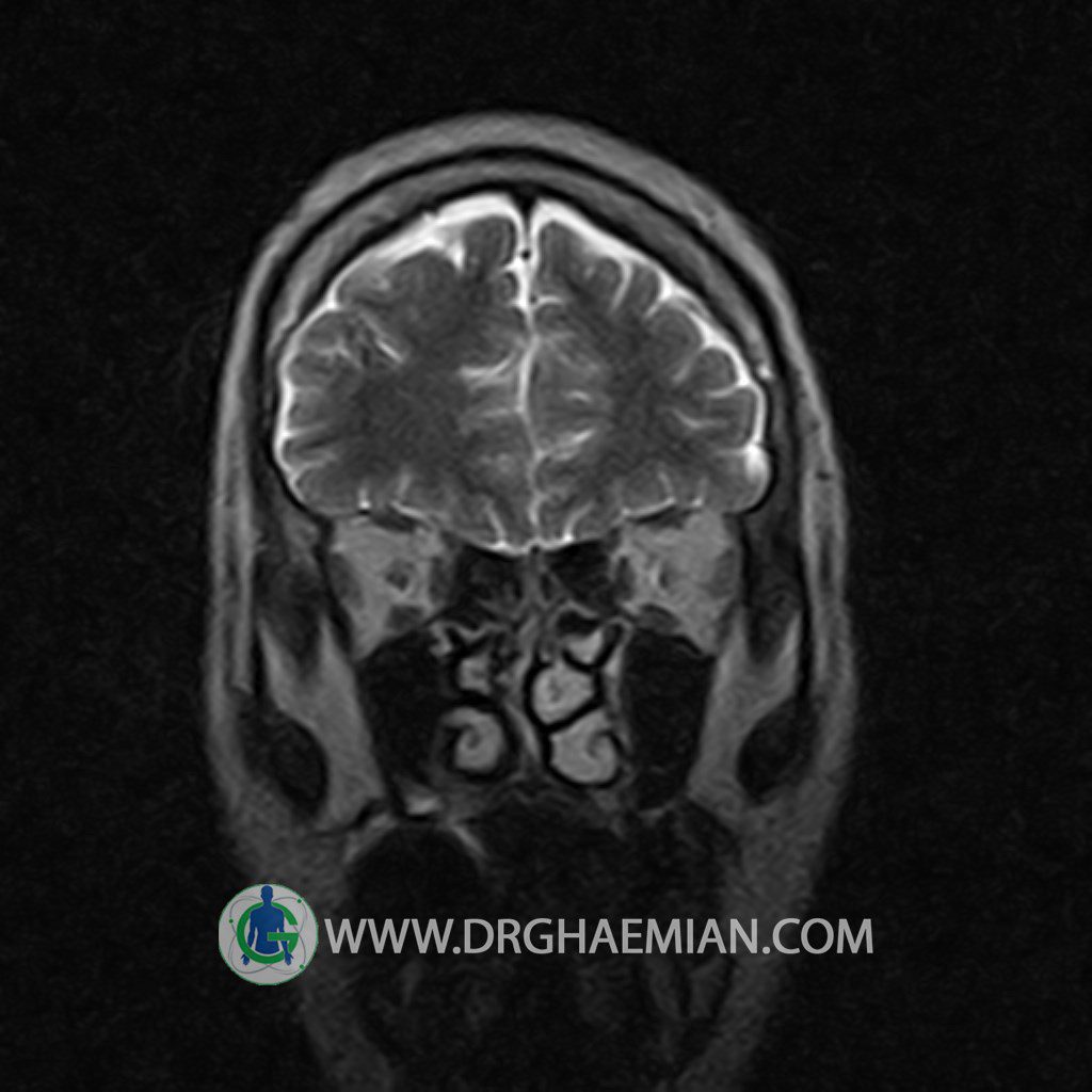
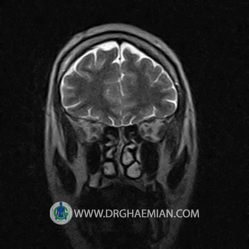
پزشکان اغلب از تصویربرداری ام آر آی برای تشخیص و درمان عارضه های پزشکی که فقط با استفاده از اشعه ایکس یا میدان مغناطیسی و امواج رادیویی قابل مشاهده است، استفاده می کنند. دستگاه ام آر آی تصاویر دقیق از ساختار های داخلی بدن ایجاد می کند. در این کیس شبهه تومور اوربیت یا همان Pseudotumor cerebri و فشار بالای دون جمجمه ایی مشاهده می شود.

گزارش پزشک:
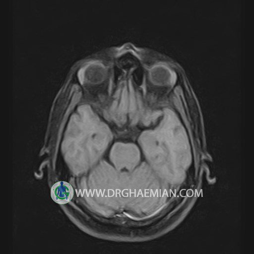
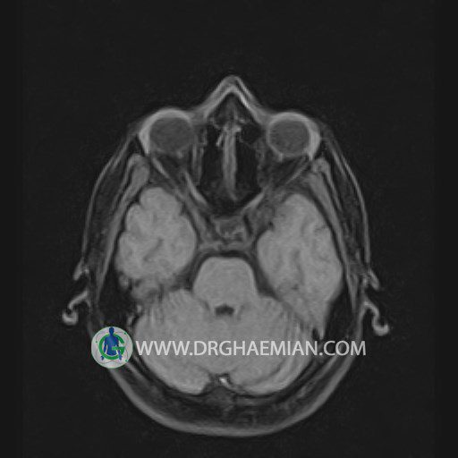
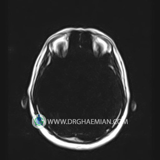
ORBIT MRI
(with and without contrast)
Technique:Axial T1 , Axial , sagittal , coronal FSE T2 , coronal T1, sagittal fat sat T2 , Axial , sagittal T1 post Gd .
REPORT :
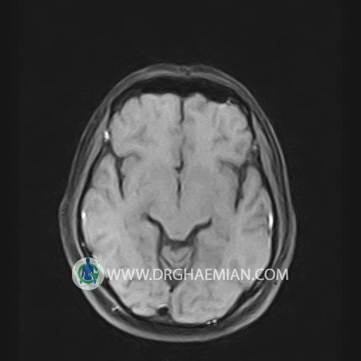
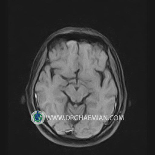
The both orbit are symmetrical and of normal size , with normal development of the orbital cones .
The bony orbital walls show a normal configuration with smooth and, sharp margins .
No foci of bone destruction , no circumscribed expansion of the bony or soft – tissue components of the orbital are evident .
The globes are symmetrical and of normal size and the ocular contents show normal signal characteristics .
The ocular walls are smooth , sharply defined , and of normal thickness .
The eye muscles are normally positioned and display normal course and width.
The retrobulbar fat, ophthalmic vein and lacrimal apparatus are unremarkable .
No seen any evidence of ocular herniation .
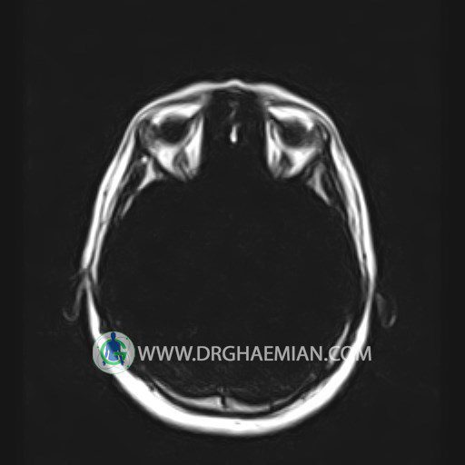
– Combination of partial empty sella & tortuosity of optic nerves is suggestive for high ICP & pseudotumor cerebri
is seen
