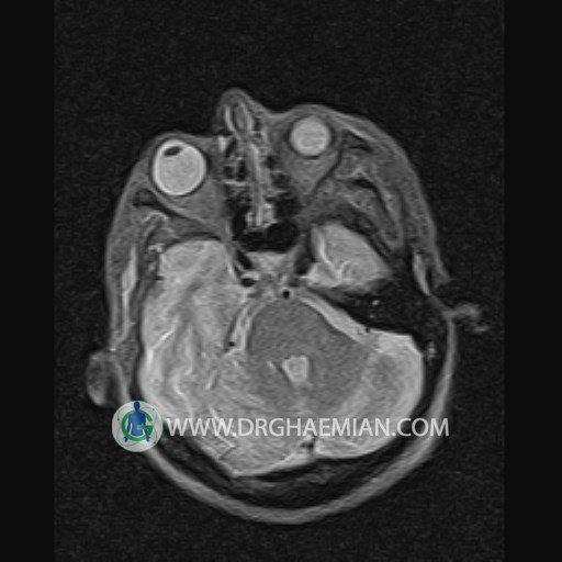
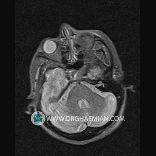
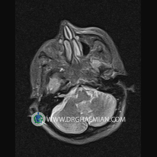
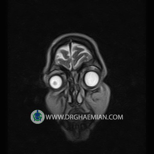
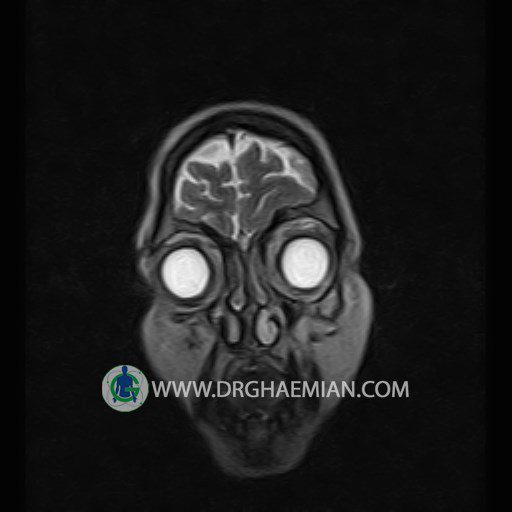
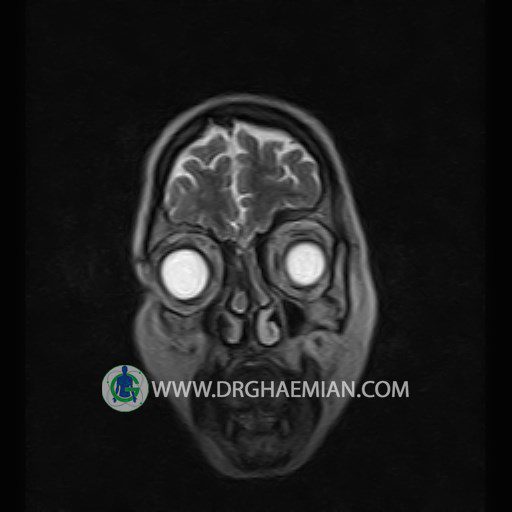
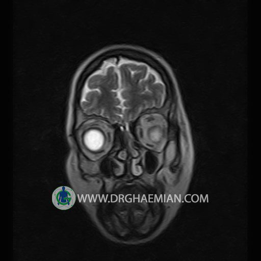
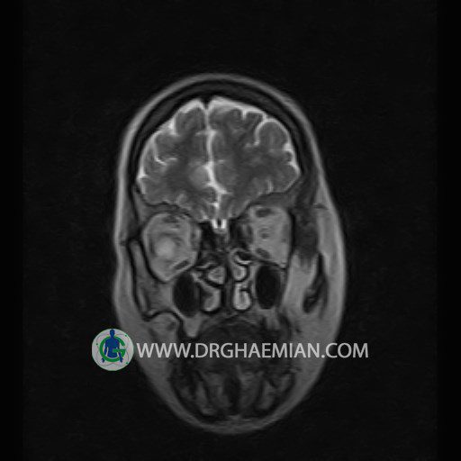
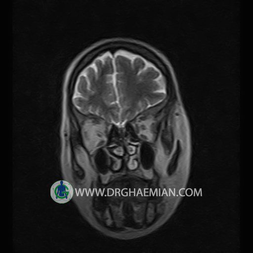
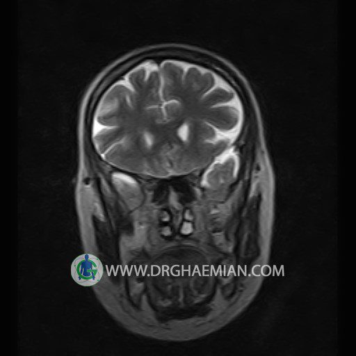
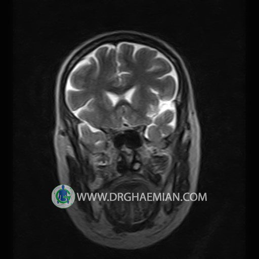
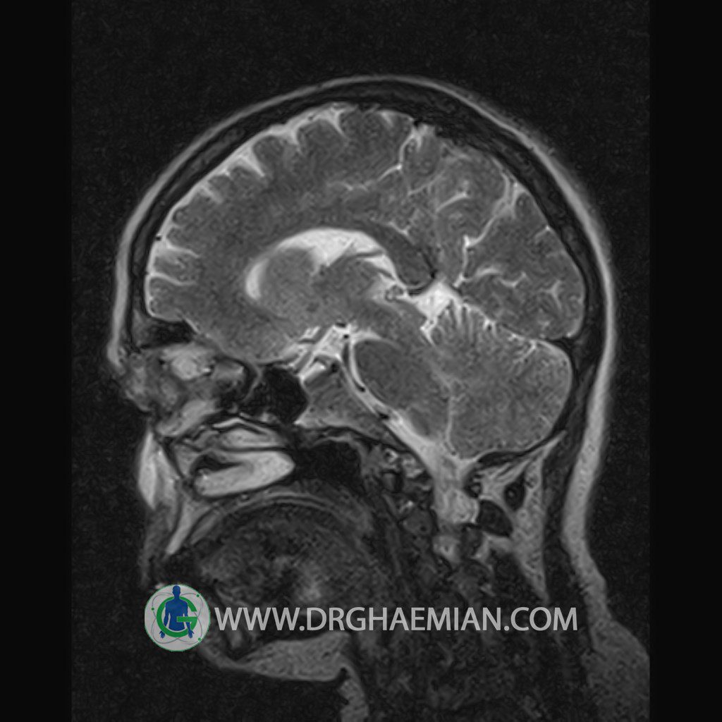
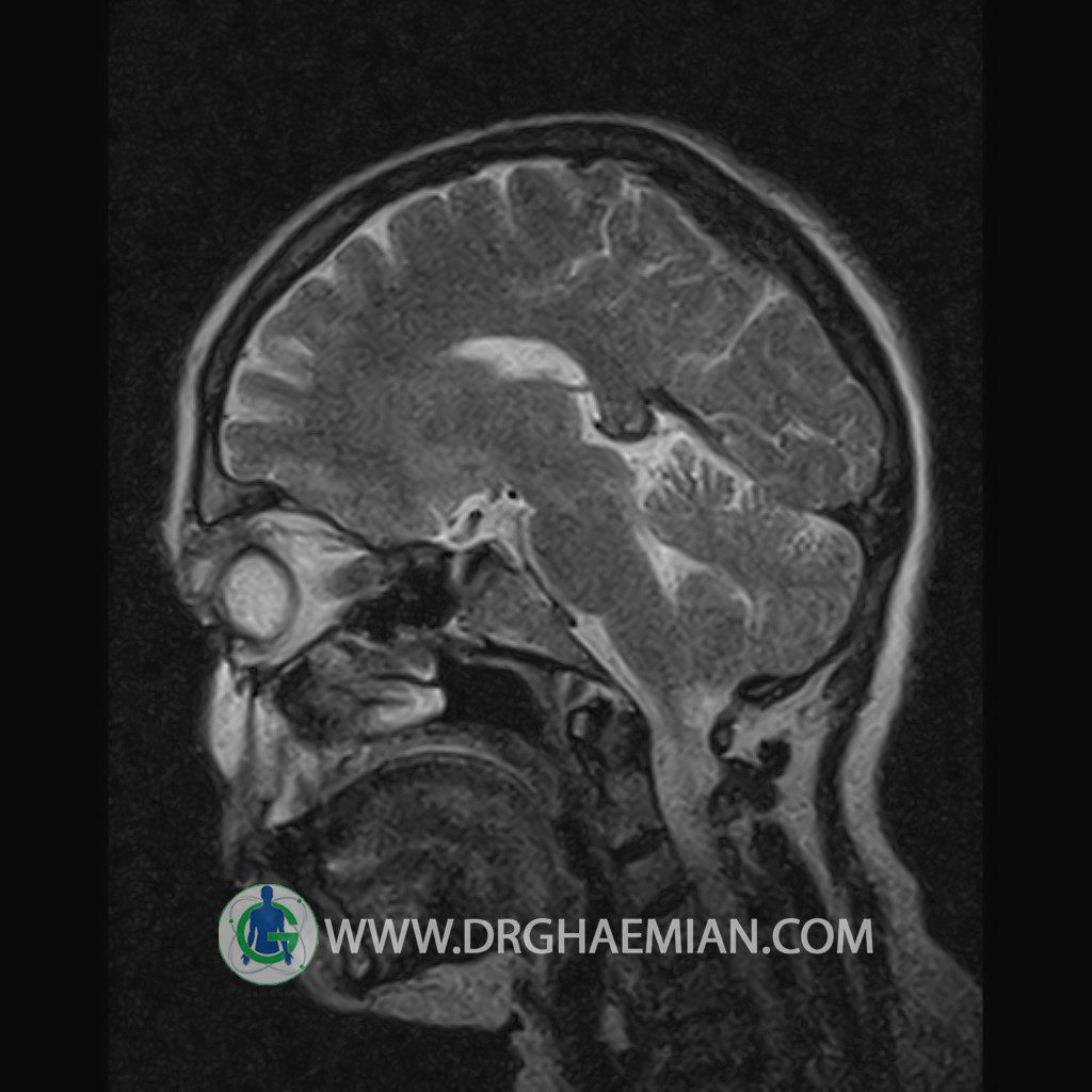
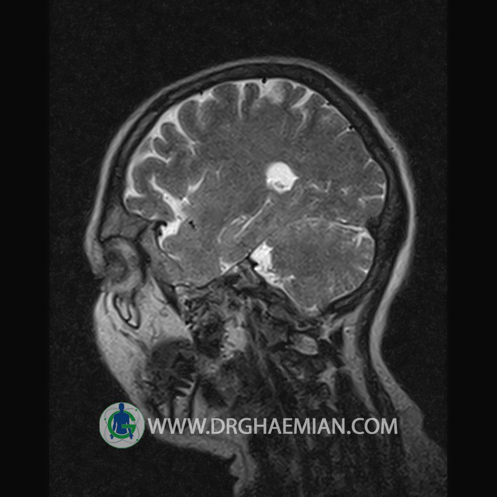
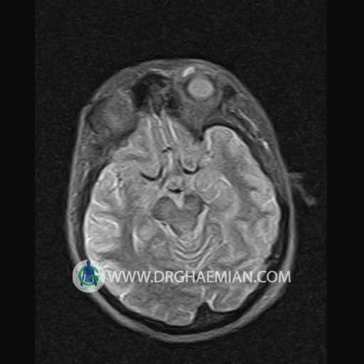
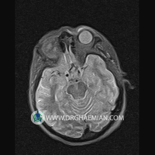
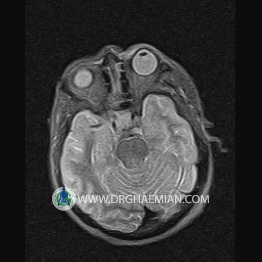
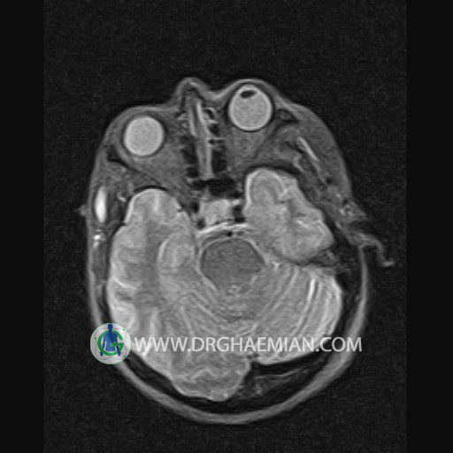
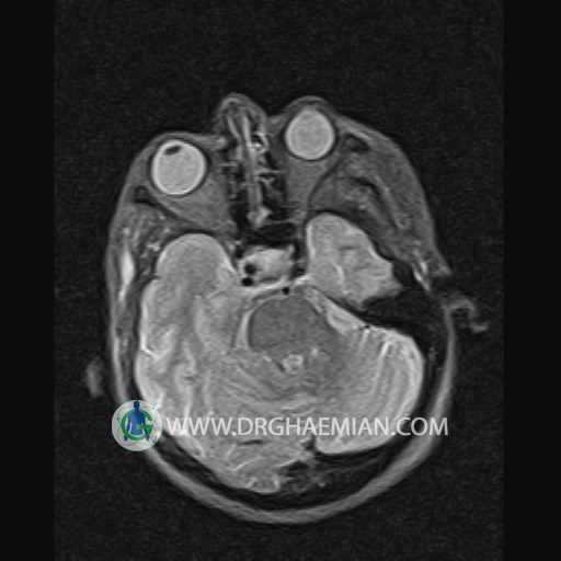
پزشکان اغلب از تصویربرداری ام آر آی برای تشخیص و درمان عارضه های پزشکی که فقط با استفاده از اشعه ایکس یا میدان مغناطیسی و امواج رادیویی قابل مشاهده است، استفاده می کنند. دستگاه ام آر آی تصاویر دقیق از ساختار های داخلی بدن ایجاد می کند. در این کیس سلولیت (عفونت بافت) در اوربیت بیمار مشاهده می شود.
گزارش پزشک:
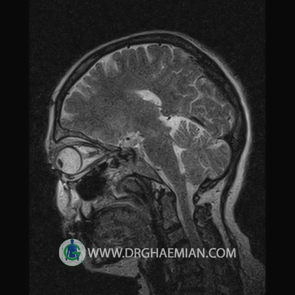
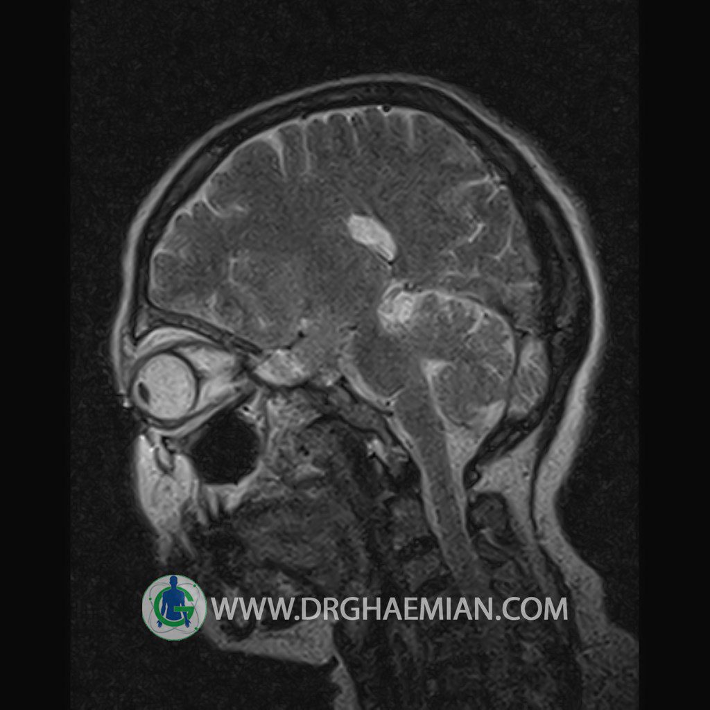
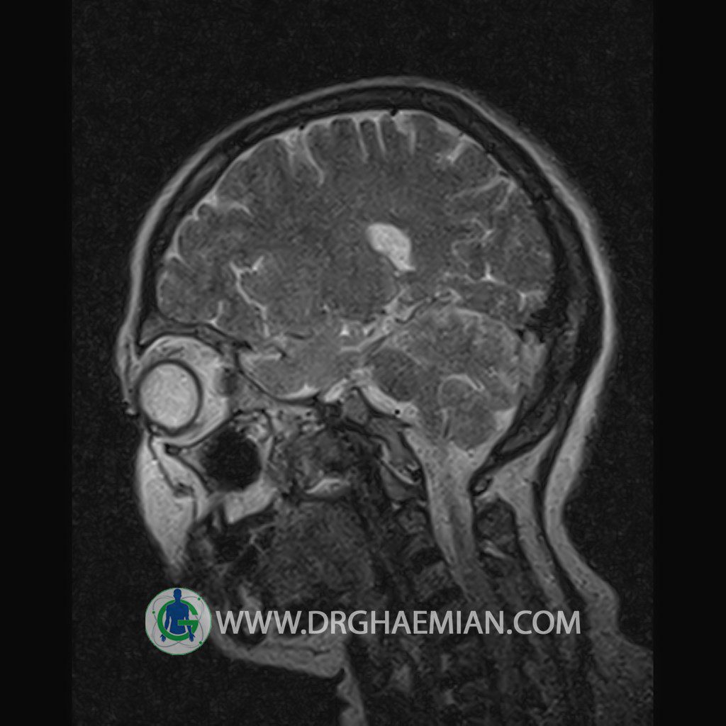
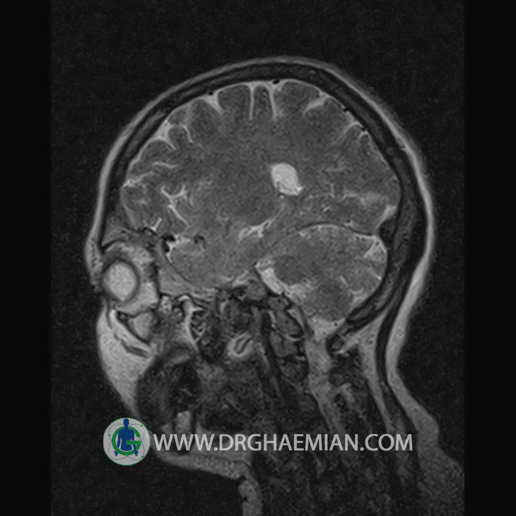
ORBIT MRI
(with and without contrast)
Technique:Axial T1 , Axial , sagittal , coronal FSE T2 , coronal T1, sagittal fat sat T2 , Axial , sagittal T1 post Gd .
REPORT :
The bony orbital walls show a normal configuration with smooth and, sharp margins .
The ocular walls are smooth , sharply defined , and of normal thickness .
The optic nerve has normal course and caliber on each side .
The eye muscles are normally positioned and display normal course and width.
The retrobulbar fat, ophthalmic vein and lacrimal apparatus are unremarkable .
Evaluable portions of the neurocranium and paranasal sinuses show no abnormalities
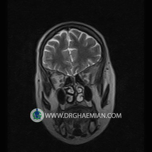
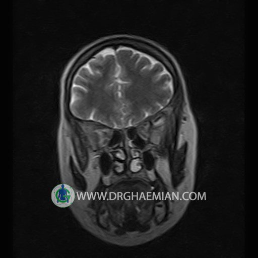
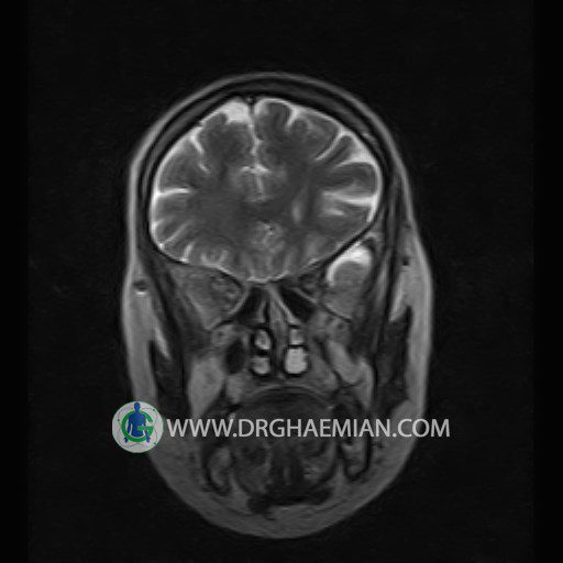
– Soft tissue swelling in anterior of left glob & in left frontal scalp with post contrast mild enhancement suggestive for cellulitis
is seen
– In comparison to previous MRI , Decreased in size of periorbital inflammation is suggestive for response to medical therapy.
