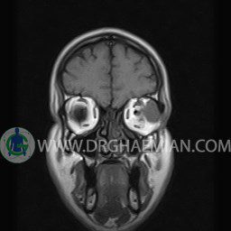
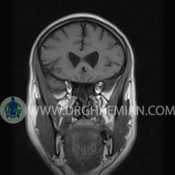
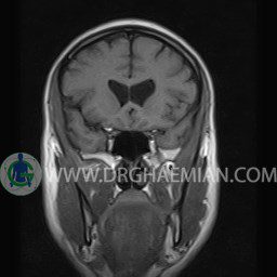
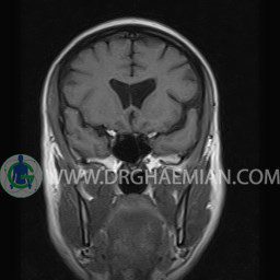
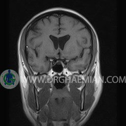
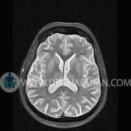
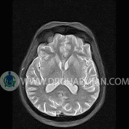
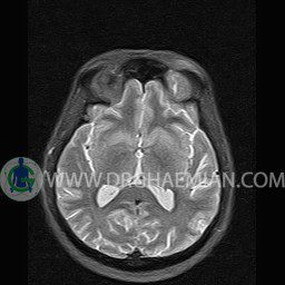
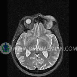
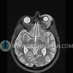
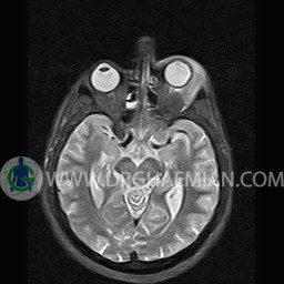
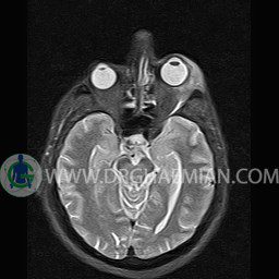
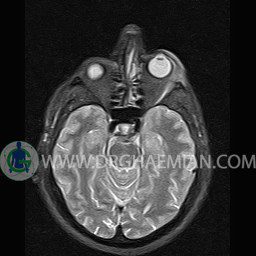
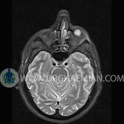
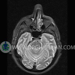
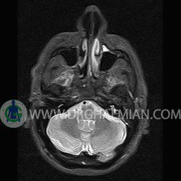
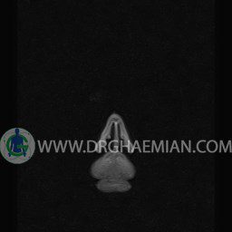
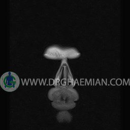
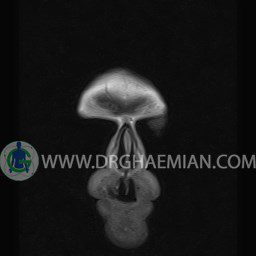
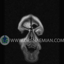
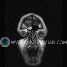
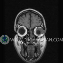
ام آر آی اوربیت با استفاده از آهنربا و امواج رادیویی تصاویری از اوربیت ها، اعصاب، عضلات و بافت های اطراف آن ایجاد می کند. ذر این کیس ضایعه نئوپلاستیک اروبیت به ابعاد mm 15 x 18 x 23 دیده می شود.

گزارش پزشک :
ORBIT MRI
(with and without contrast)
Technique: Axial T1 , Axial , sagittal , coronal FSE T2 , coronal T1, sagittal fat sat T2 , Axial , sagittal T1 post Gd .
REPORT :
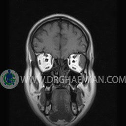
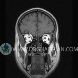
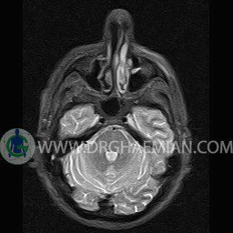
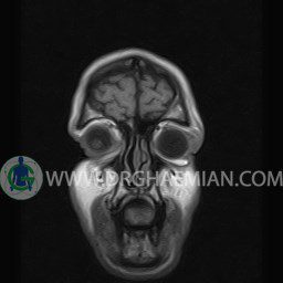
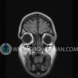
The right orbit are symmetrical and of normal size , with normal development of the orbital cones .
The globes are symmetrical and of normal size and the ocular contents show normal signal characteristics .
The ocular walls are smooth , sharply defined , and of normal thickness .
The optic nerve has normal course and caliber on each side .
The eye muscles are normally positioned and display normal course and width.
The retrobulbar fat, ophthalmic vein and lacrimal apparatus are unremarkable .
Evaluable portions of the neurocranium and paranasal sinuses show no abnormalities .
– A well – defined mass lesion ( 15 x 18 x 23 mm ) in left orbit with left proptosis & medial deviation of left lateral rectus , attached to lateral wall of orbit & with post contrast enhancement suggestive for neoplasic lesion & metastasis is seen
