
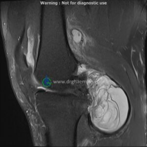
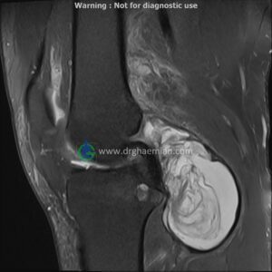
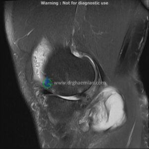
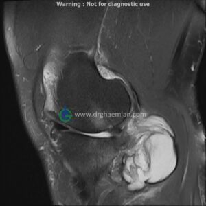
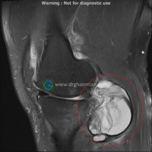
ام آر آی زانو یک روش تصویربرداری پیشرفته است که با استفاده از امواج مغناطیسی و بدون تابش پرتو، تصاویری دقیق از ساختارهای داخلی زانو، مانند رباطها، غضروفها و استخوانها، تهیه میکند تا مشکلاتی مانند آسیبهای رباطی، آسیبهای غضروفی و مشکلات مفصلی را شناسایی کند. در این کیس کیست سینوویال… دیده میشود

گزارش پزشک
LEFT KNEE MRI
(Without contrast)
Siemens MRI ( magnetom altea 1.5 tesla )
Technique: Sagittal T1, Axial T2 fat sat, coronal & sagittal PD fat sat .
The bones comprising the knee joint are normal in configuration.
The cortical bone has normal thickness.
Lateral meniscus displays normal configuration.
MCL & LCL are intacted.
Patellar ligamentum and quadriceps tendon are normal in shape and signal intensity .
– Knee joint effusion with soft tissue swelling around the knee
– Full thickness cartilage loss with bone reaction in medial condyle of femur & in medial plateau of tibia (grade 4 chondromalacia)
– Medial meniscus extrusion with maceration
– Nonvisualized ACL & buckling of PCL suggestive for old tearing of ACL with mucoid degeneration
– Synovial cyst (45×75mm) in posterior of knee
are seen.