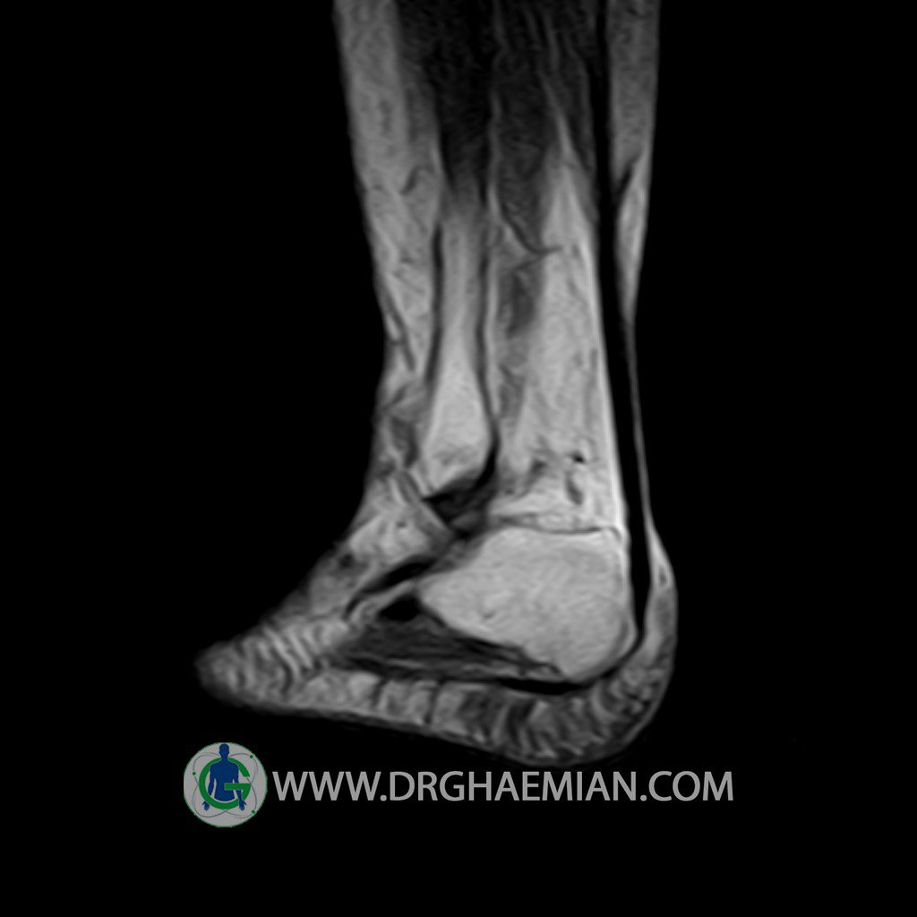
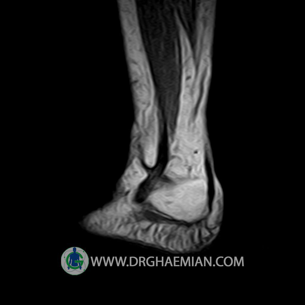
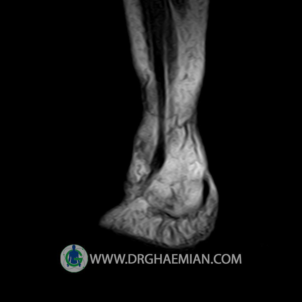
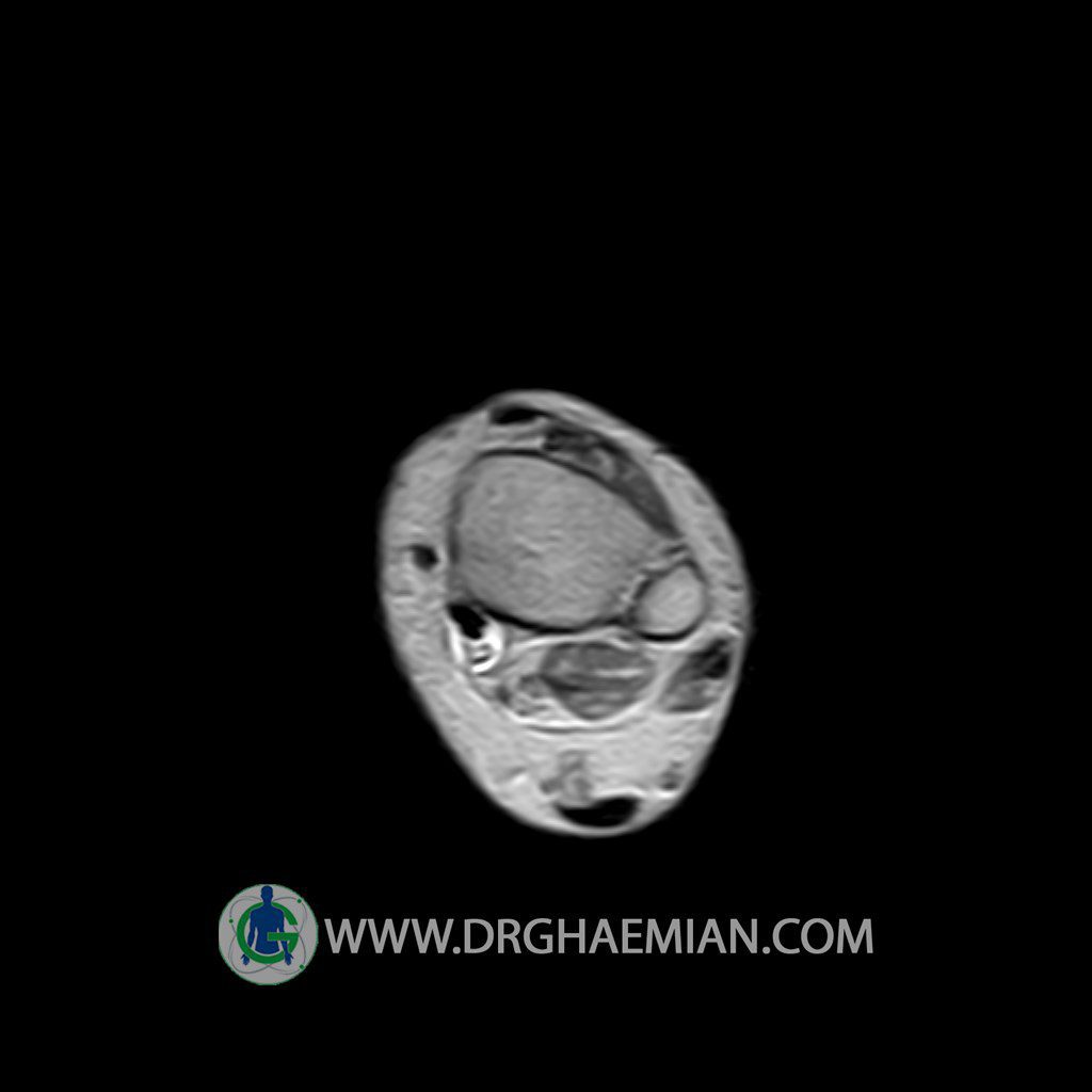
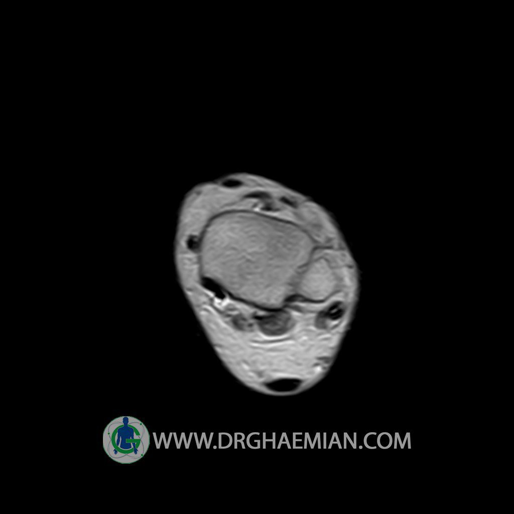
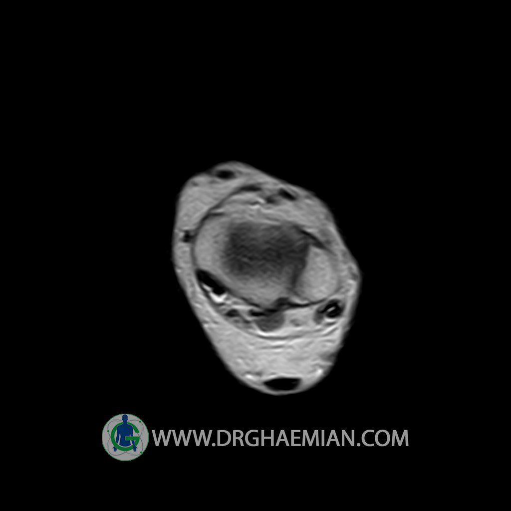
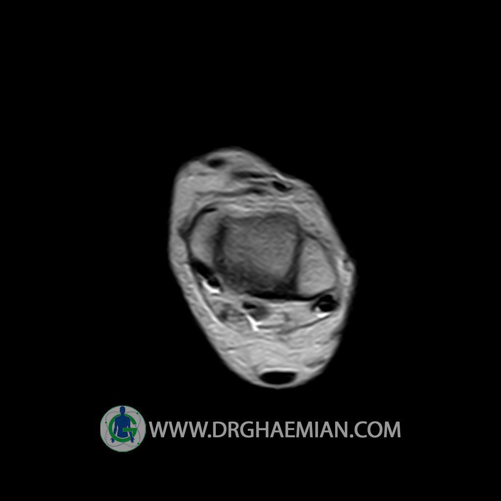
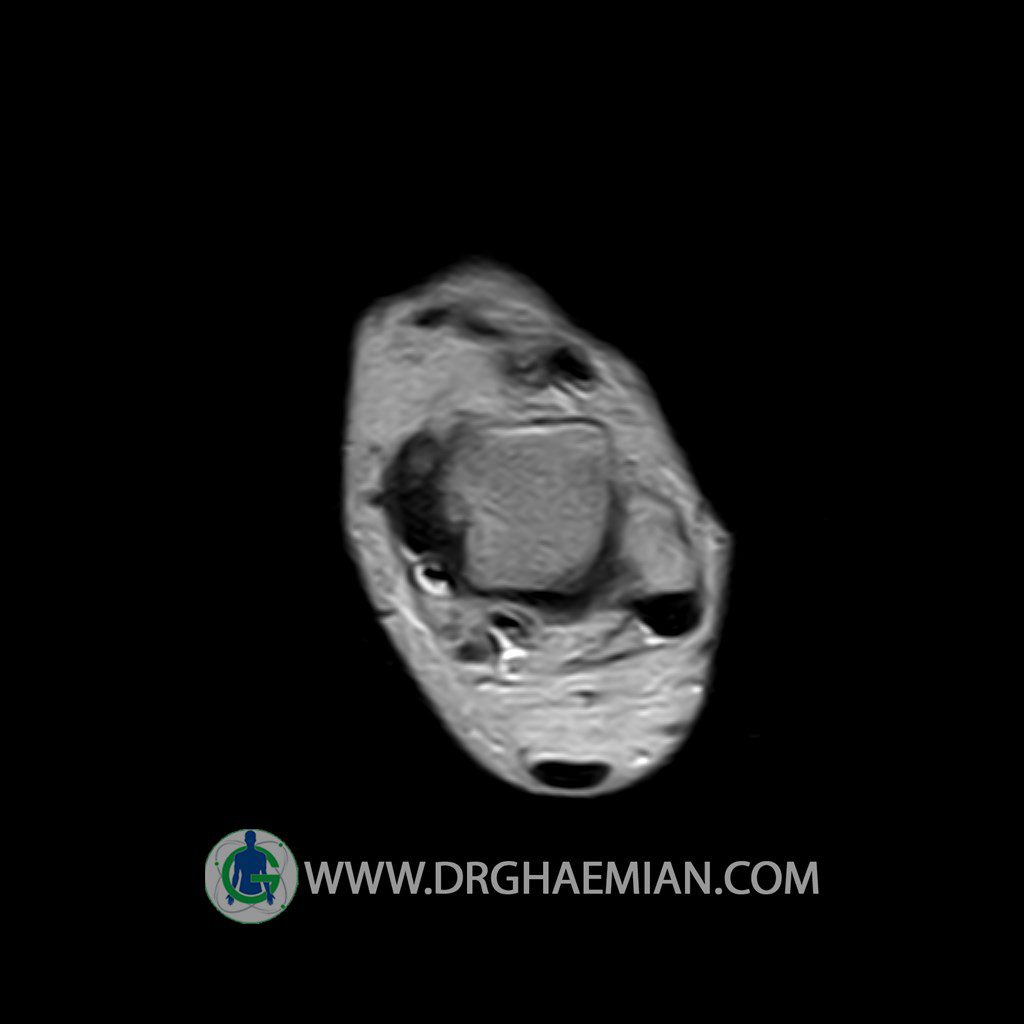
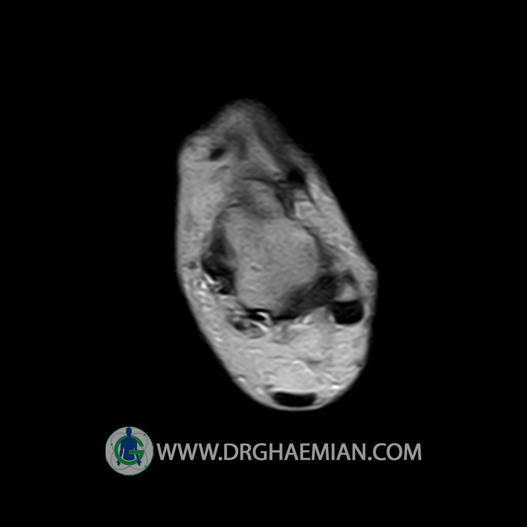
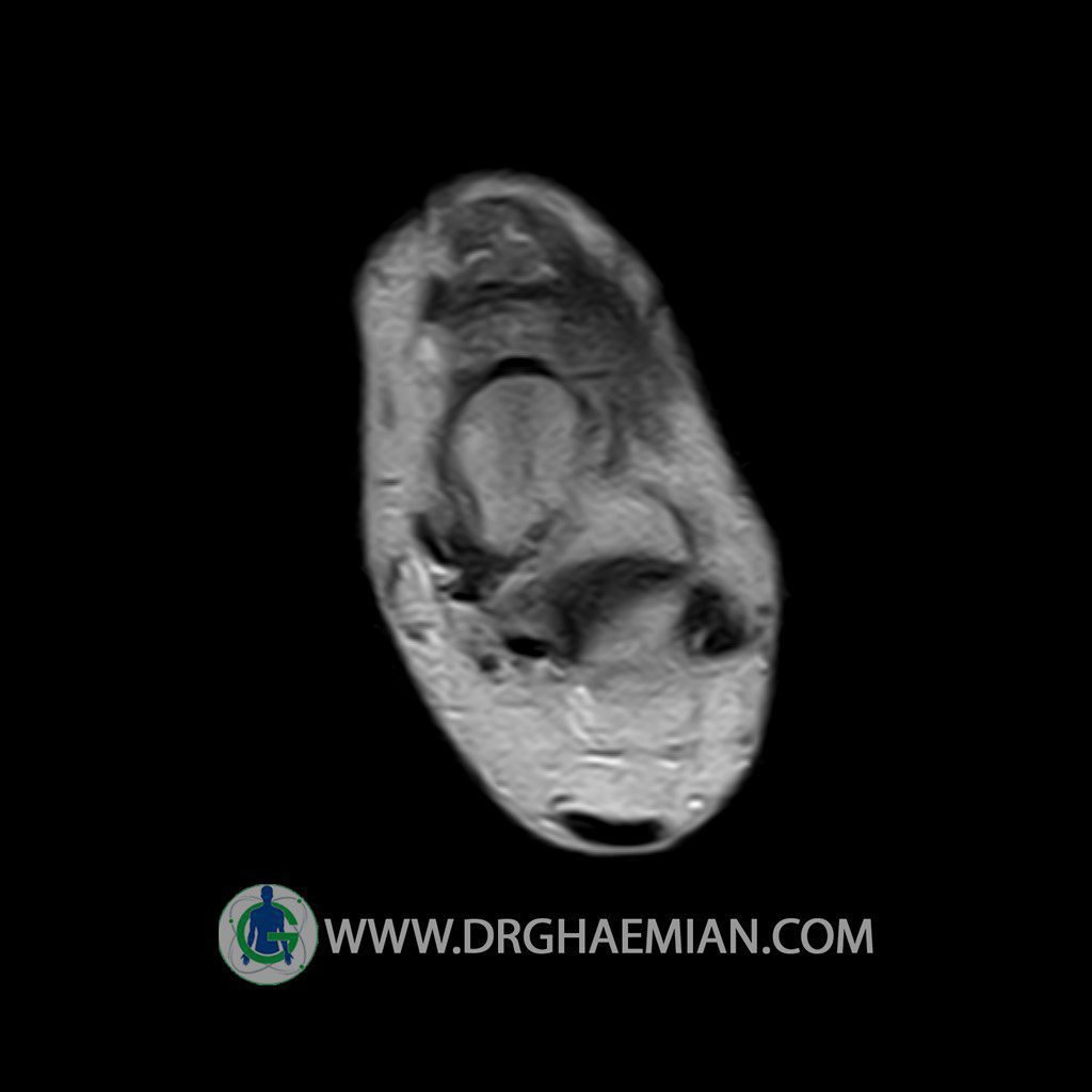
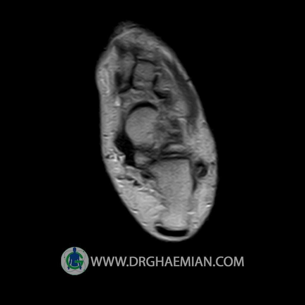
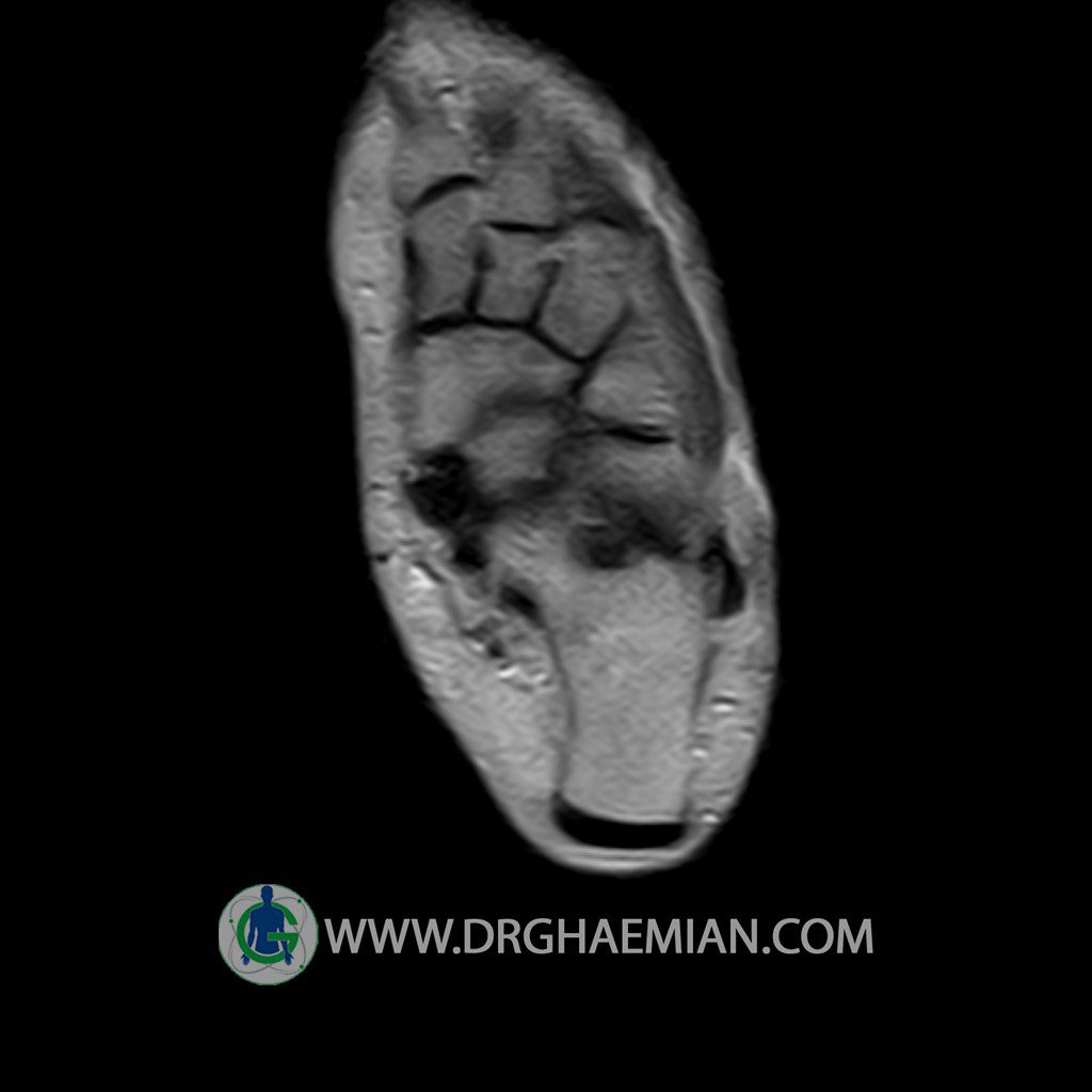
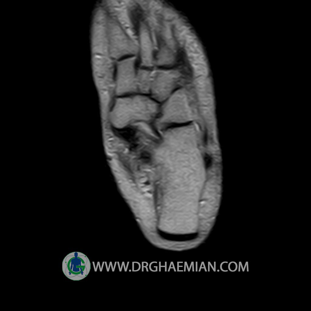
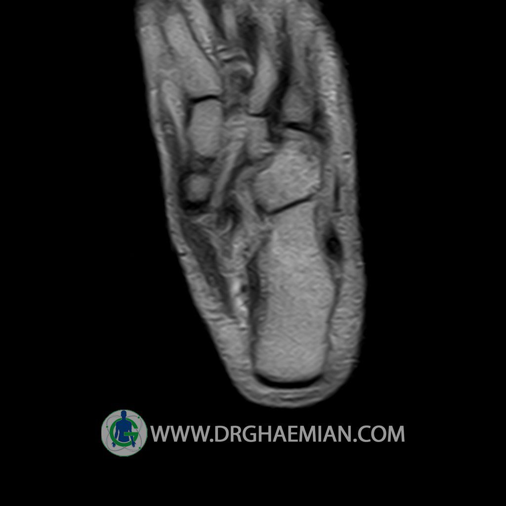
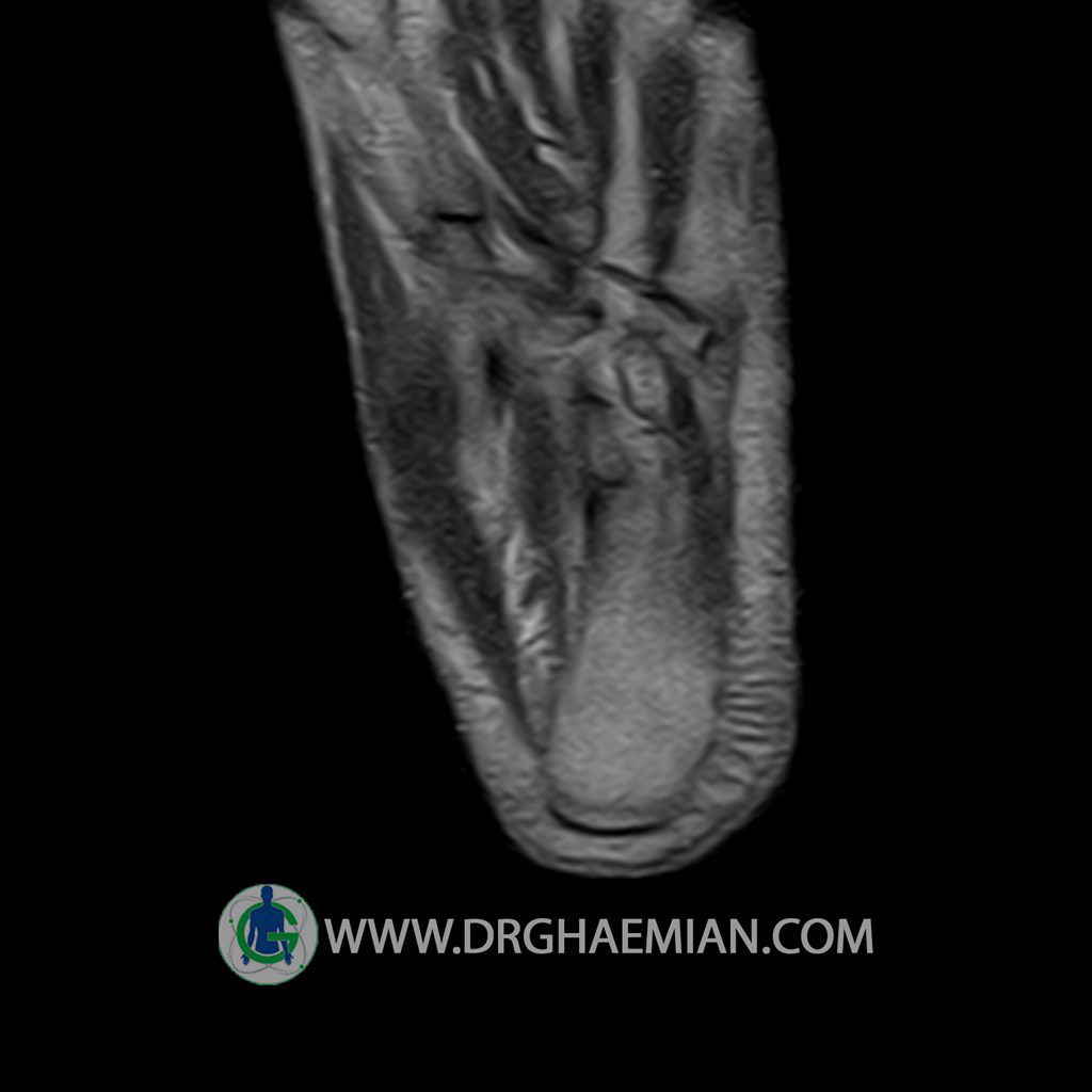
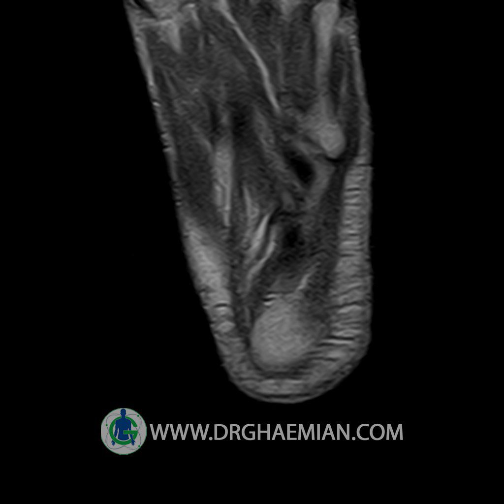
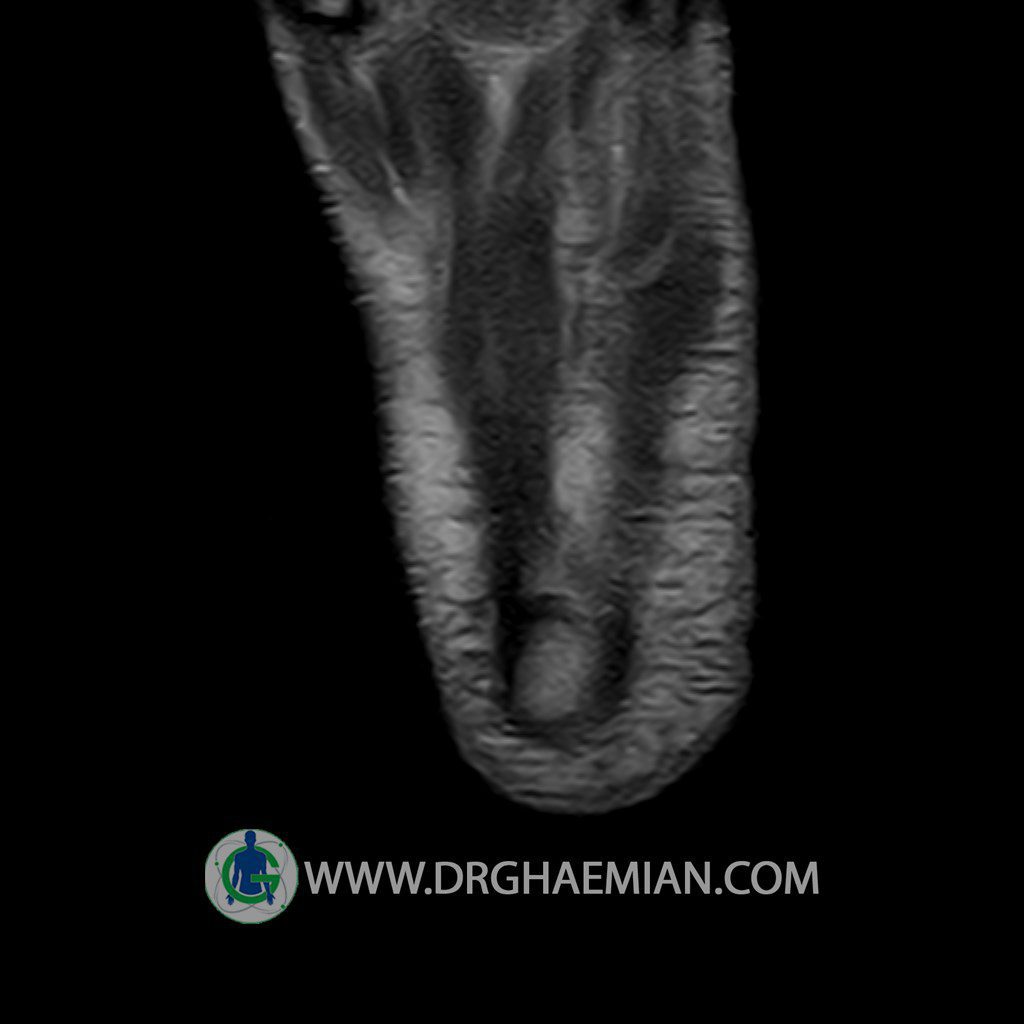
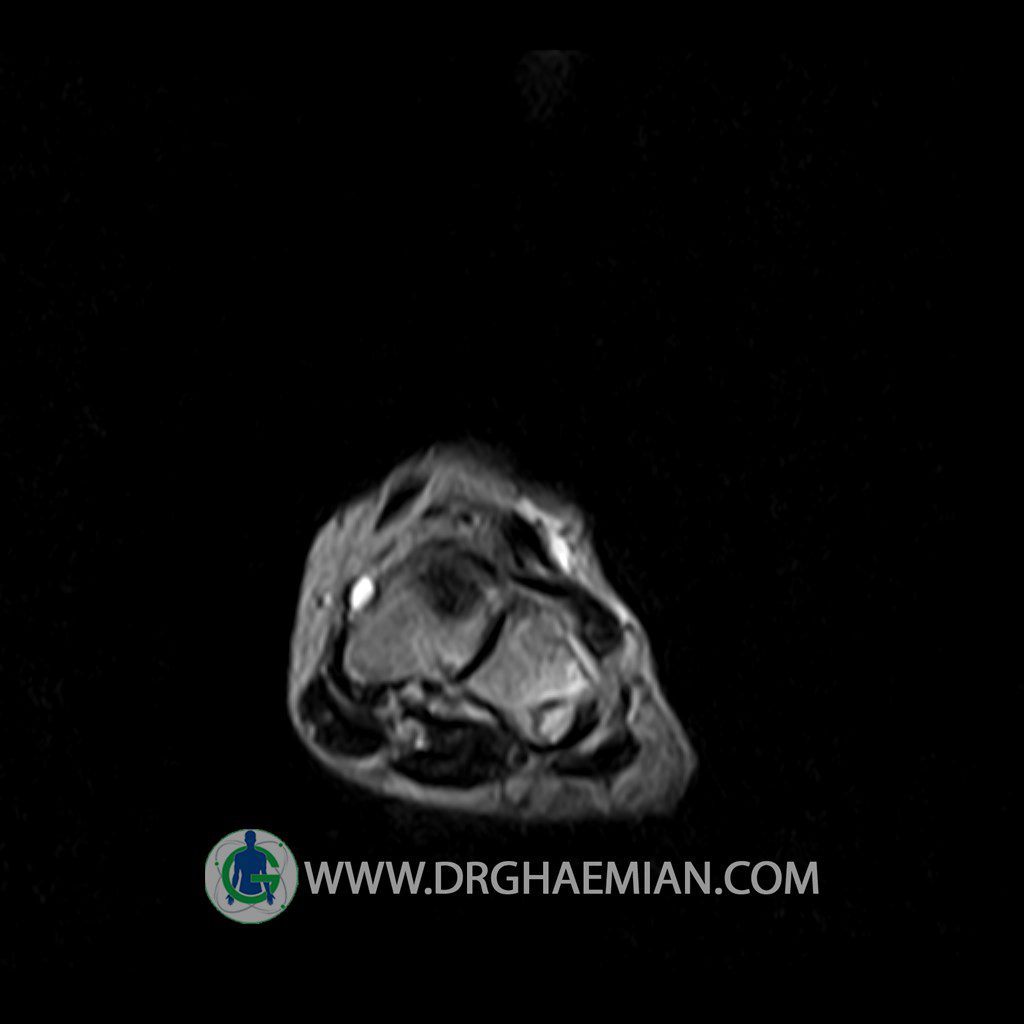
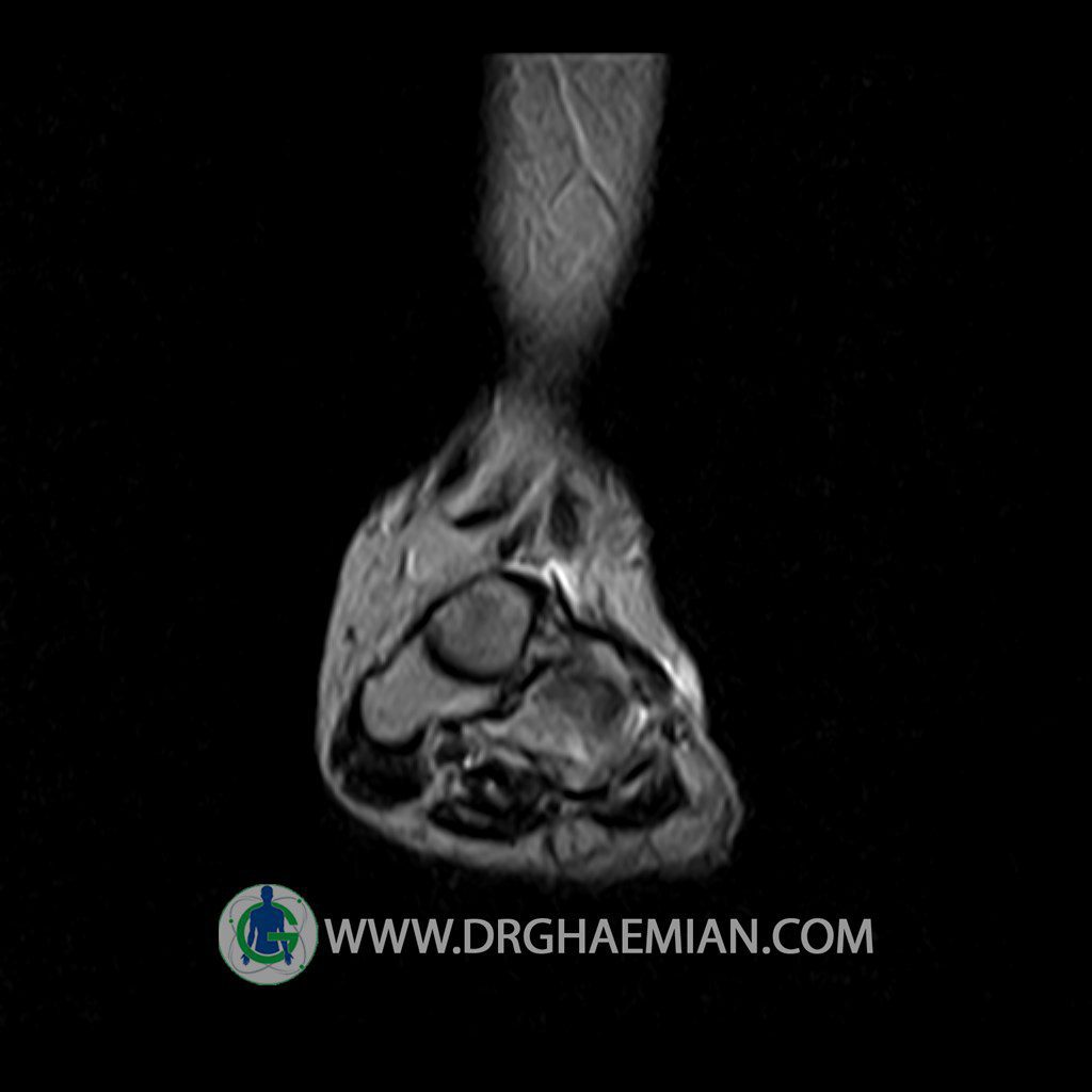
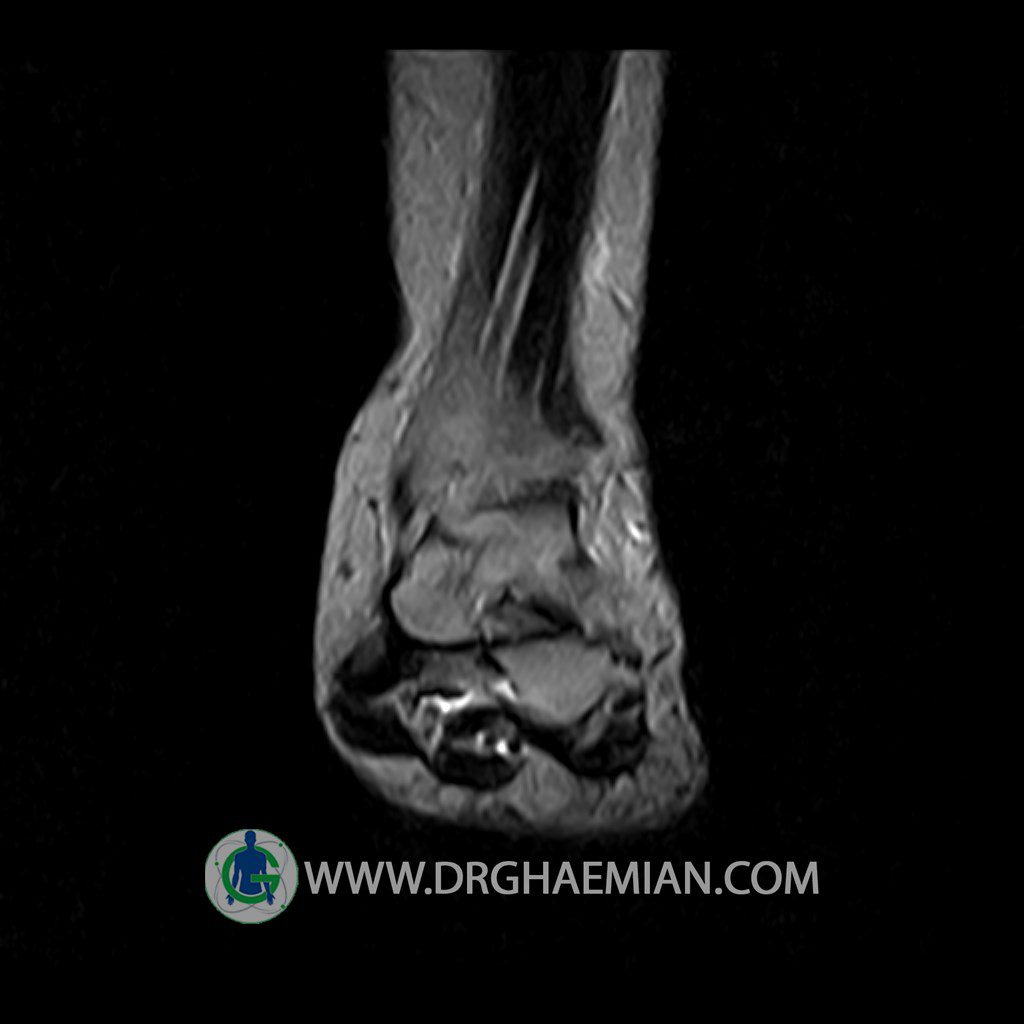
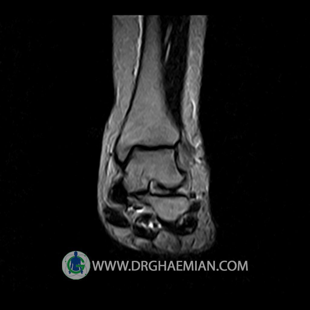
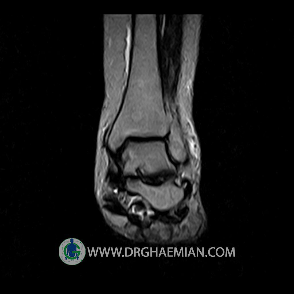
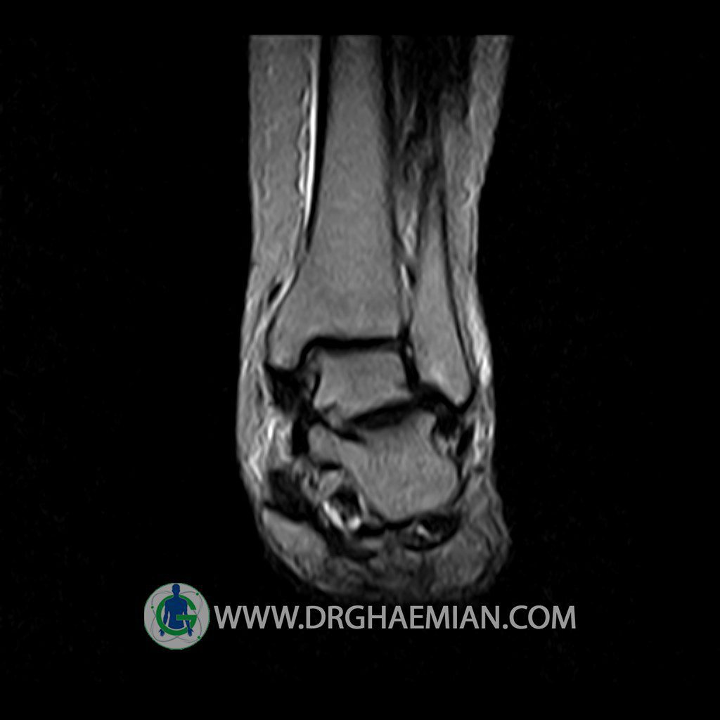
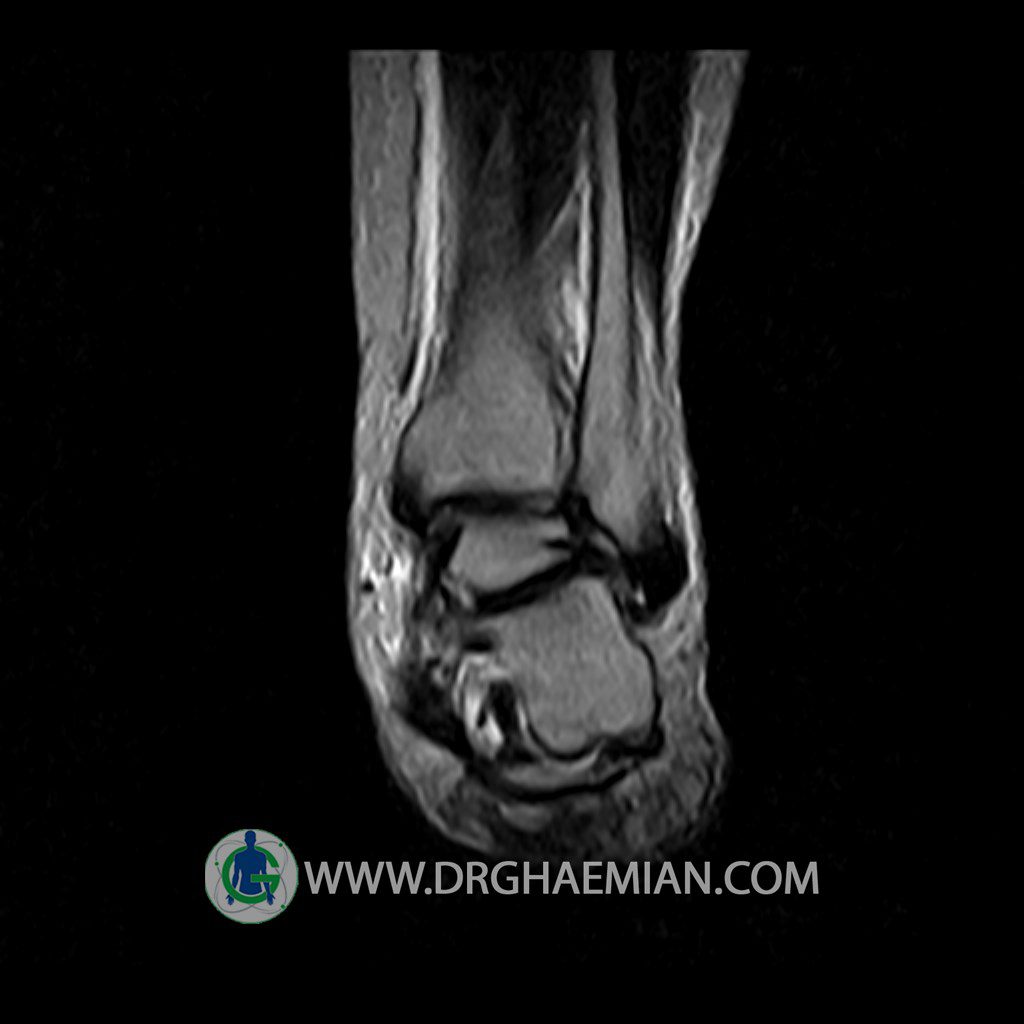
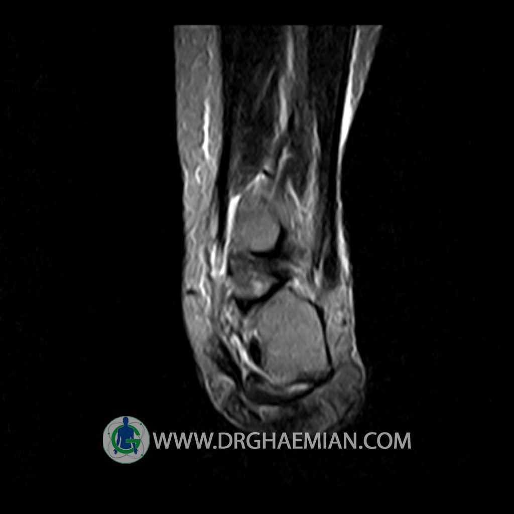
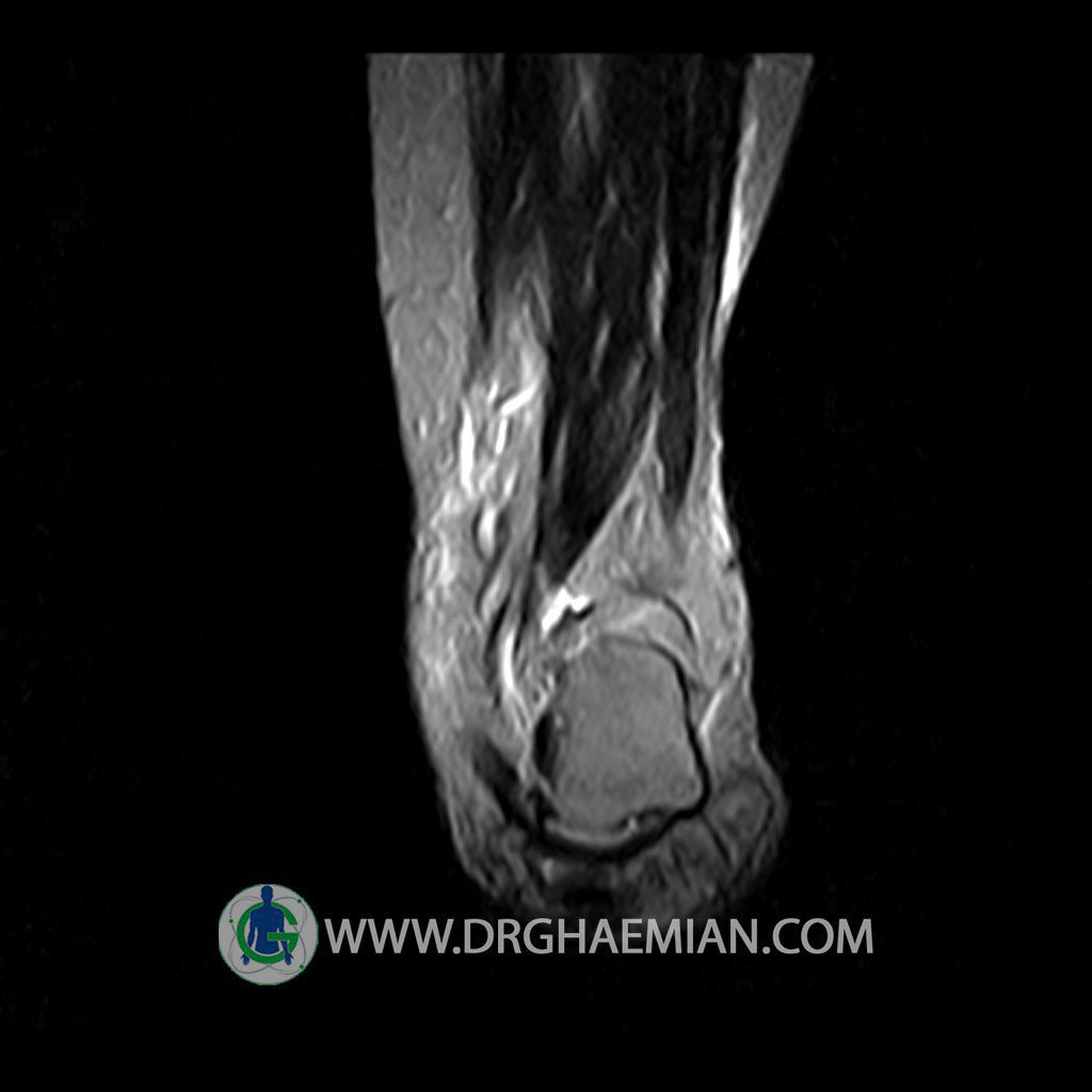
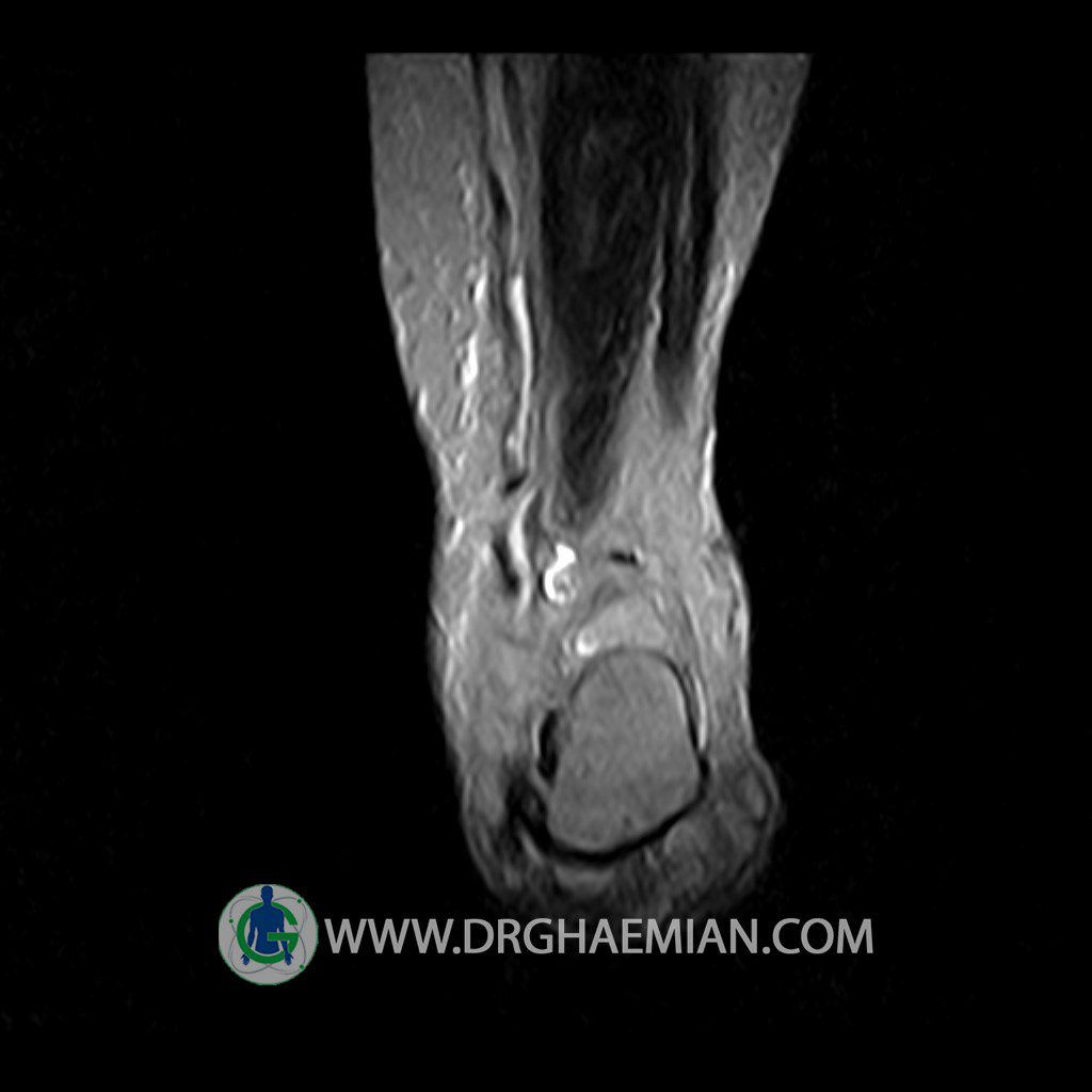
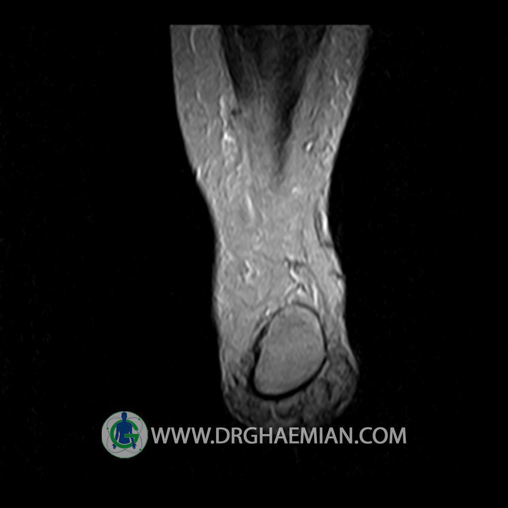
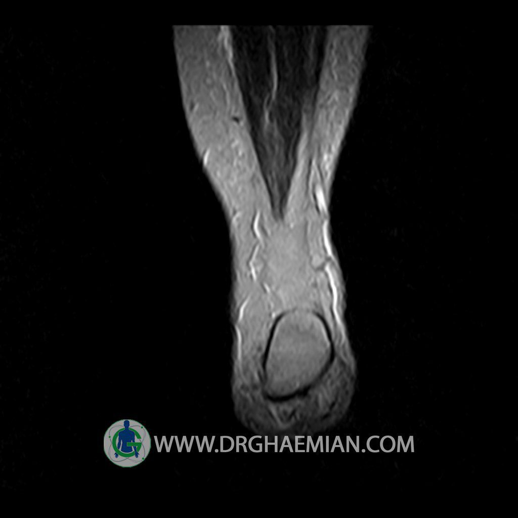
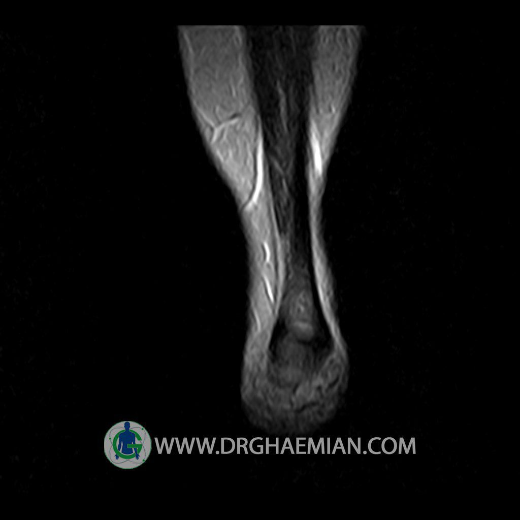
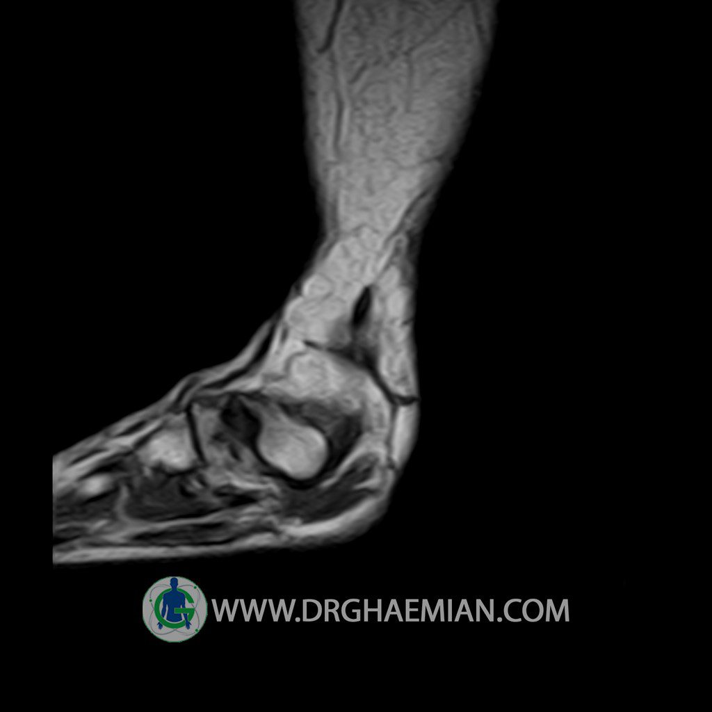
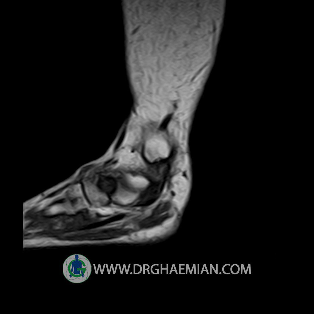
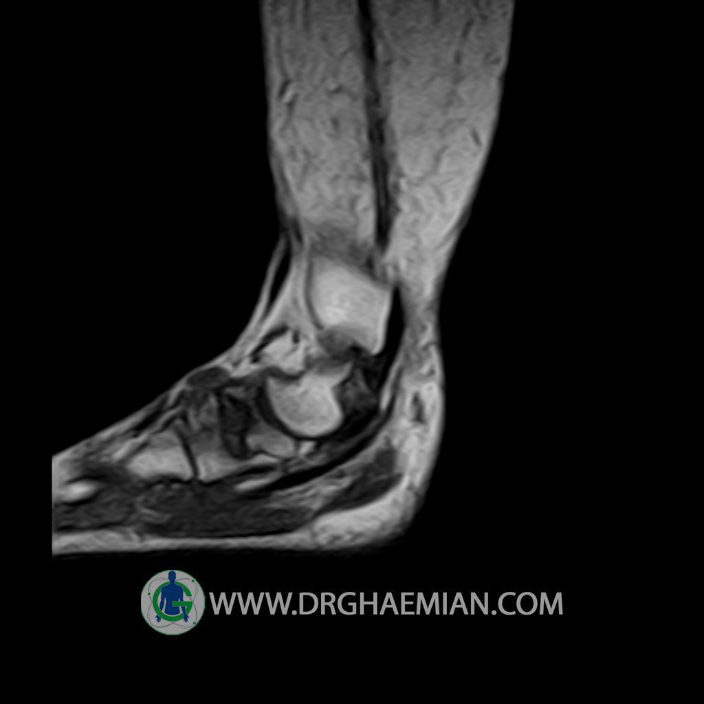
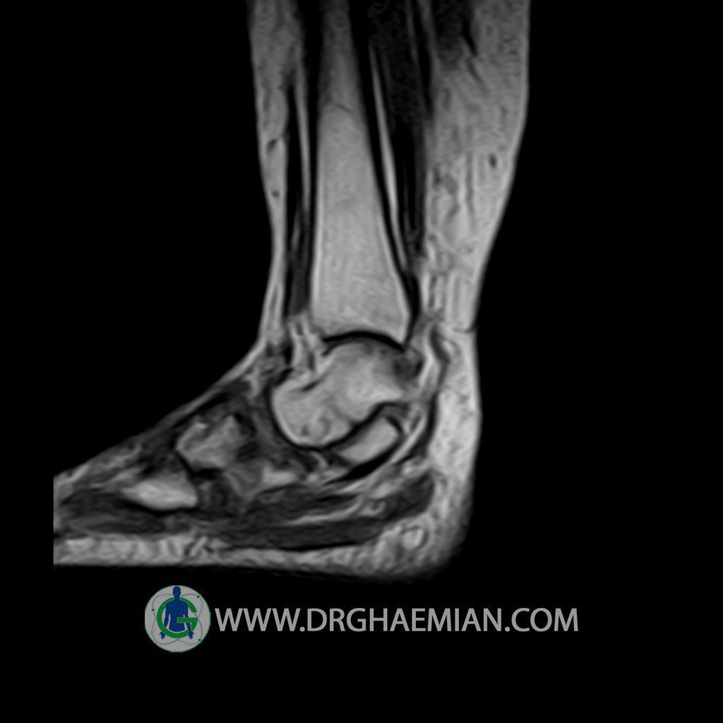
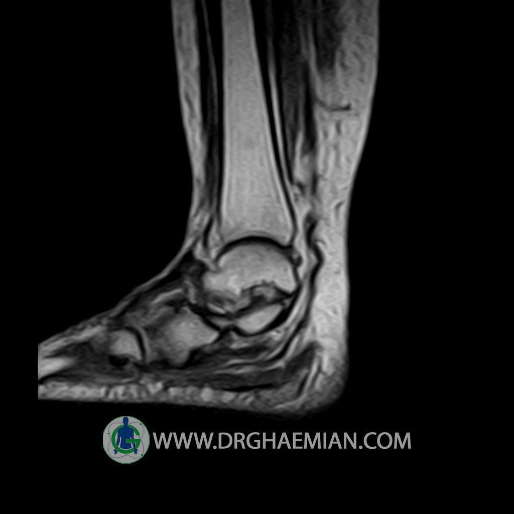
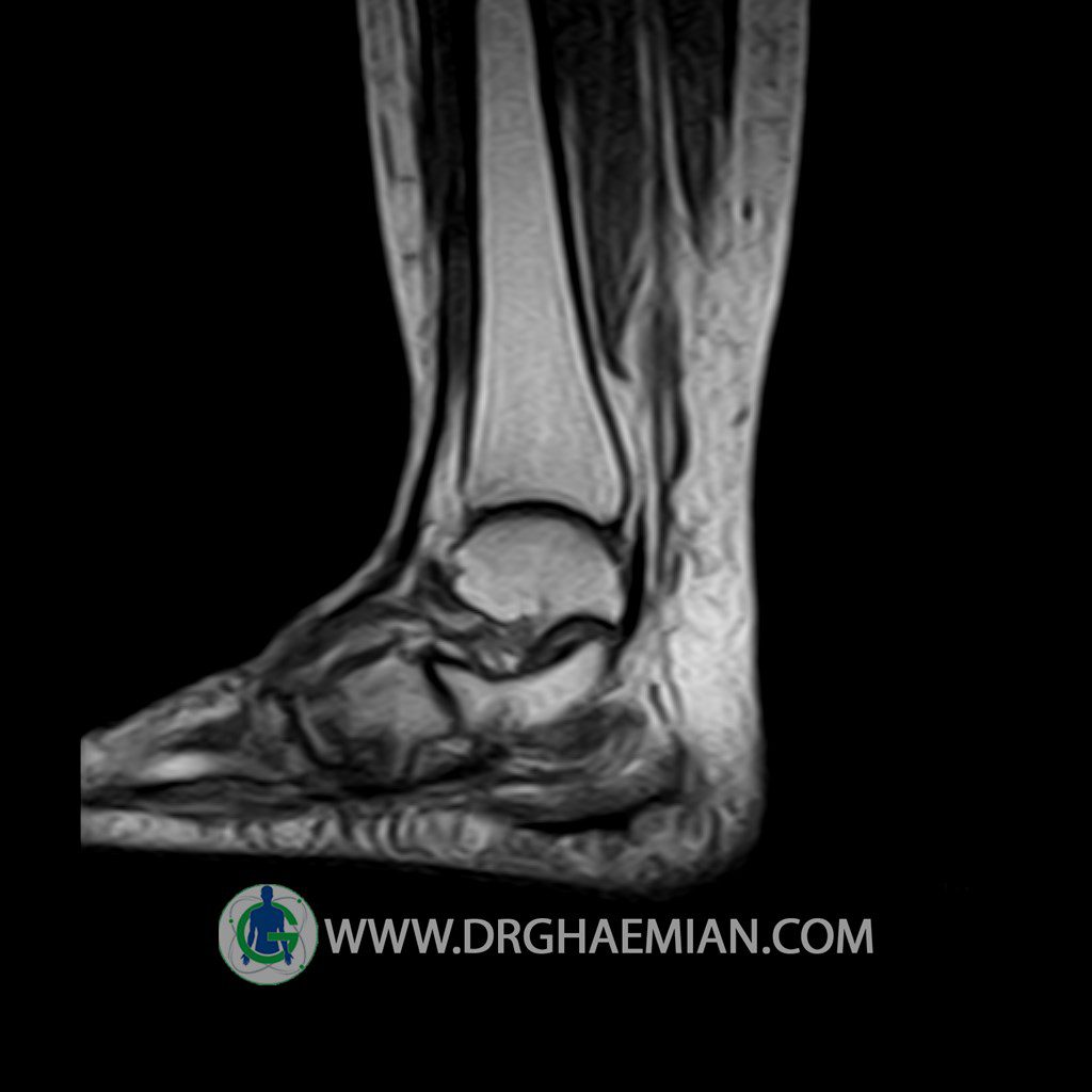
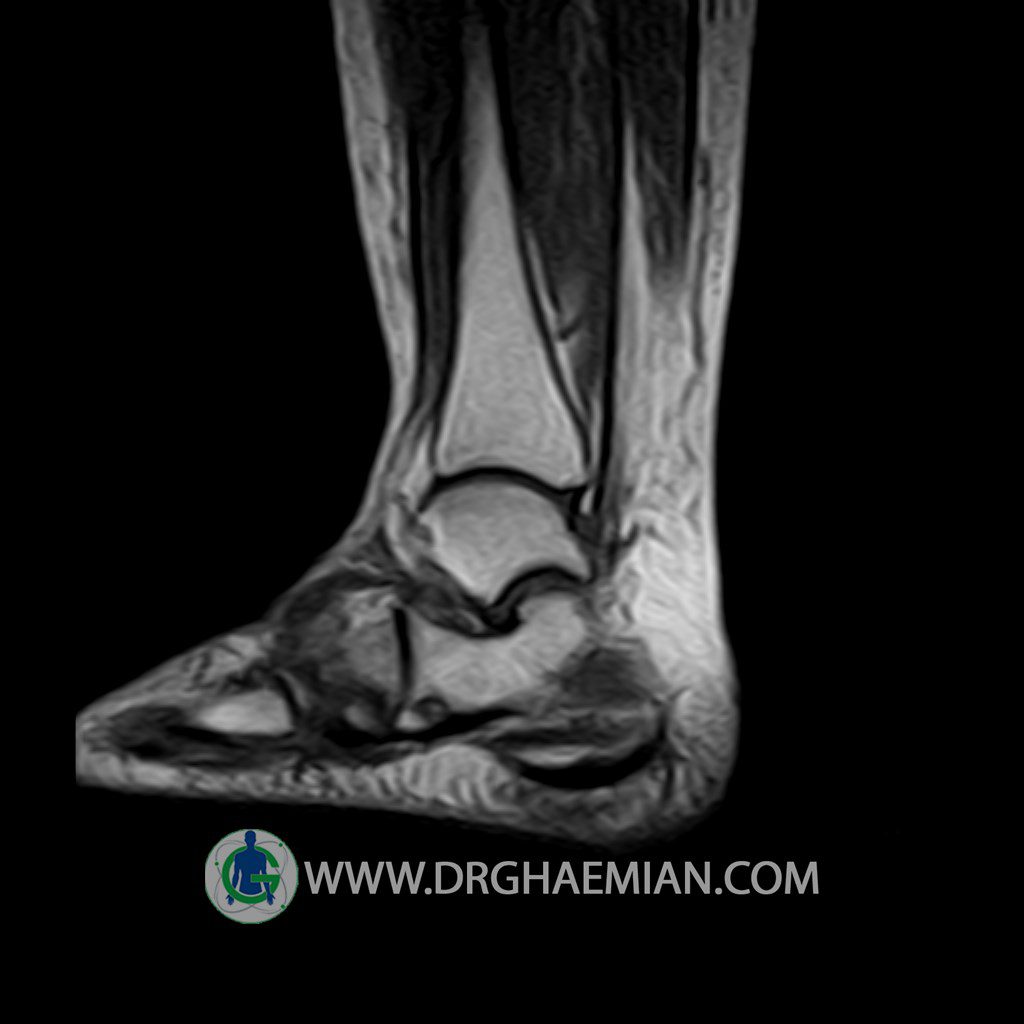
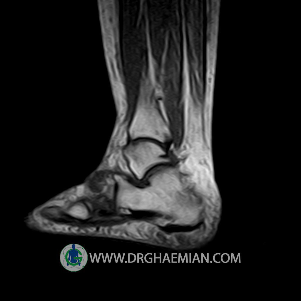
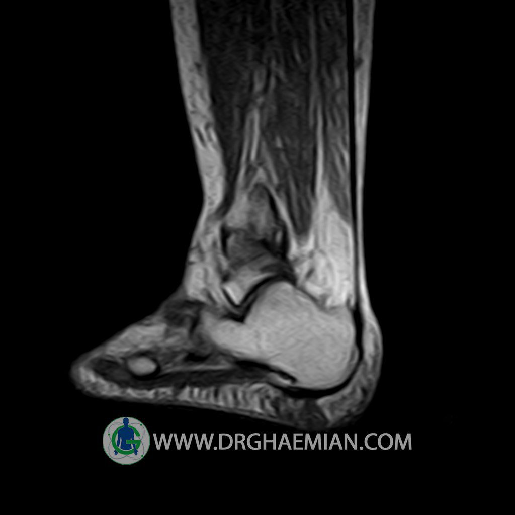
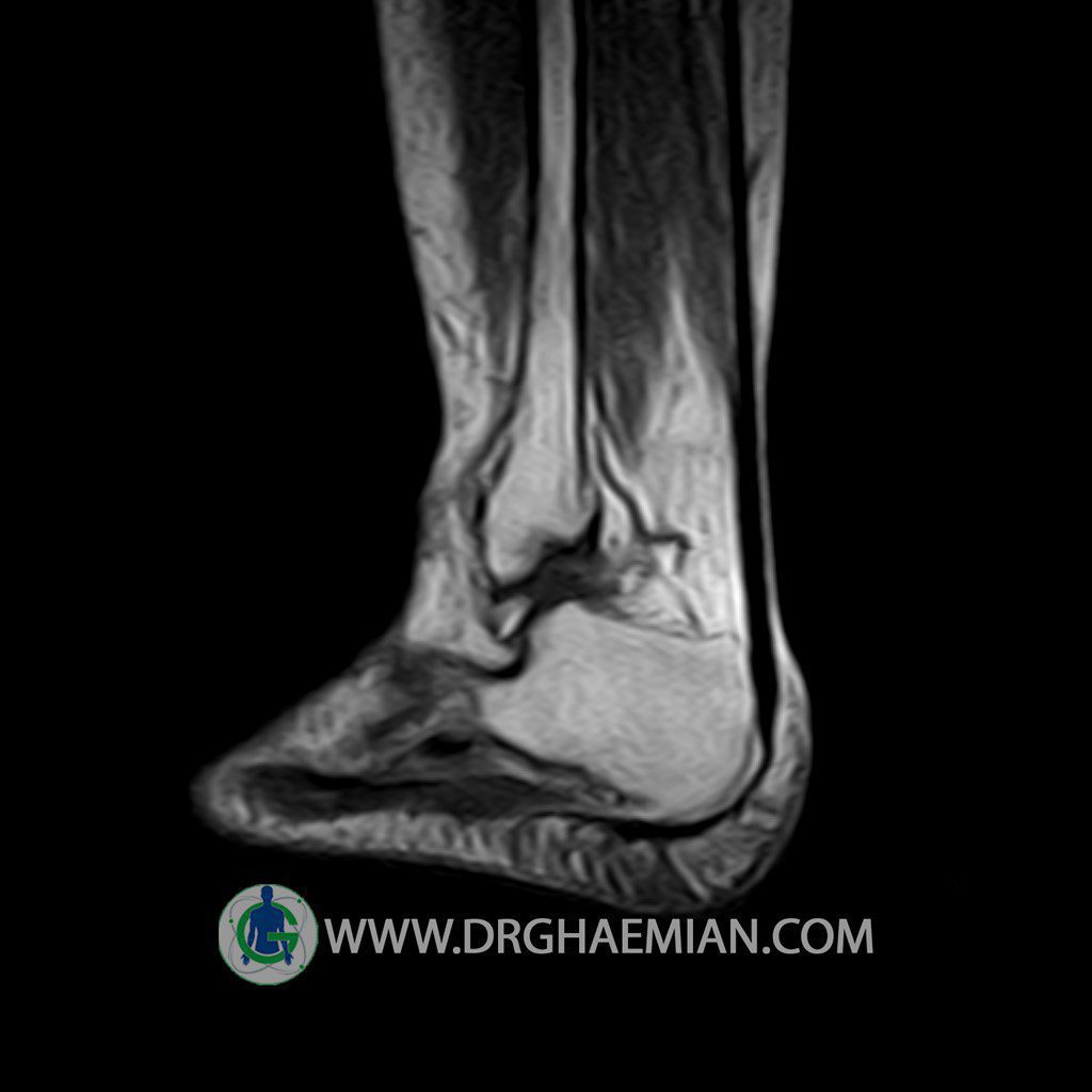
پزشکان اغلب از تصویربرداری ام آر آی برای تشخیص و درمان عارضه های پزشکی که فقط با استفاده از اشعه ایکس یا میدان مغناطیسی و امواج رادیویی قابل مشاهده است، استفاده می کنند. دستگاه ام آر آی تصاویر دقیق از ساختار های داخلی بدن ایجاد می کند. در این کیس زائده خوش خیم مچ پا، التهاب در اطراف زانو، پارگی در رباط، التهاب در اطرف مچ دیده می شود.


گزارش پزشک:
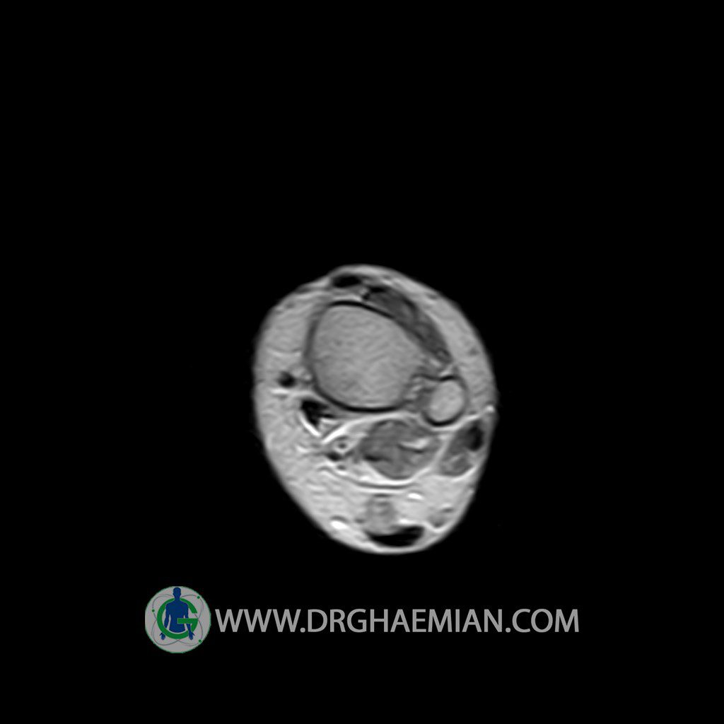
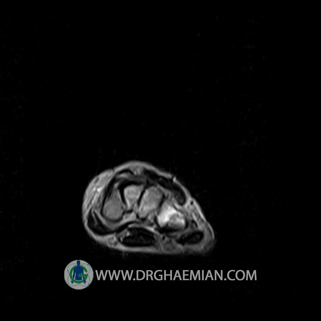
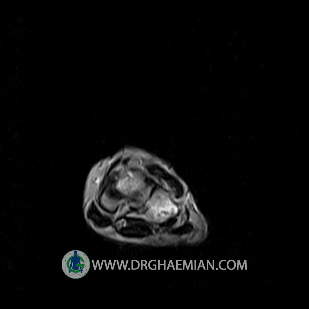
LEFT KNEE MRI
(Without contrast)
Technique: Sagittal T1, T2 , Axial GE , coronal & sagittal fatsat .
The bones comprising the knee joint are normal in signal and configuration.
The cortical bone has normal thickness.
Bone marrow signal is normal .
The hyaline cartilage covering patella, femoral condyles and tibial plateau shows normal signal and thickness.
Medial and lateral meniscus displays normal configuration .
ACL , PCL ,MCL & LCL are intact.
Soft tissues surrounding the knee joint are unremarkable .
Patellar ligamentum and quadriceps tendon are normal in shape and signal intensity .
– mild soft tissue swelling around the knee
– radial tearing in P.H. of medial meniscus with meniscal extrusion
– Focal signal change with well – defined borders in mid portion of diatal metaphysis of femur suggestive for benign bone lesion
are seen
– Soft tissue swelling around the ankle
– Signal change in cuboid suggestive for bone bruise with adjacent soft tissue swelling
are seen
