ام آر آی کمر از طریق انرژی آهنربا های قوی تصاویری از قسمت پایین ستون فقرات (گودی کمر) ایجاد می کند. این نوع تصویربراری از اشعه ایکس استفاده نمی کند.
برخی از ام آر آی های کمر نیازمند رنگی مخصوص (داروی کنتراست) است. در اکثر مواقع این رنگ مخصوص توسط سرمی یا آمپول از دست یا بازوی شما تزریق می شود. این رنگ مخصوص باعث می شود رادیولوژیست برخی از قسمت ها را شفاف تر ببیند.
تصویربرداری ام آر آی بدون درد است. شما باید بی حرکت دراز بکشید چرا که حرکت کردن میتواند تصویر ام آر آی را محو کرده و خطا ایجاد کند.
دلایل تجویز ام آر آی کمر
شما ممکن است نیاز به ام آر آی قسمت پایینی ستون فقرات (کمر) داشته باشید اگر:
- درد لگن یا کمر داشته باشید که بعد از درمان بهتر نشود.
- ضعف، کرخی، و دیگر نشانه ها در پا که نه بهتر می شود نه وخیمتر
اگر شما متبلا به موارد زیر هستید پزشک ممکن است ام آر آی کمر تجویز کند:
- درد کمر و تب
- نقایص مادرزادی قسمت پایین ستون فقرات
- آسیب یا صدمه به قسمت پایینی ستون فقرات
- درد کمر به همراه سابقه یا نشانه های سرطان
- ام اس
- مشکلات در کنترل یا خالی کردن مثانه
- بیرون زدگی دیسک کمر
[ngg src=”galleries” ids=”2″ display=”basic_thumbnail” thumbnail_crop=”0″]
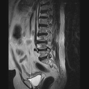
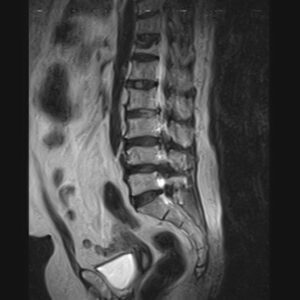
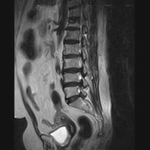
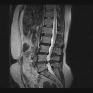
REPORT:
The imaged soft tissues show no abnormalities .
The visualized cord and filum terminalis are normal .
The conus medullaris terminates normally at L1.
Paravertebral stripe is normal in shape and signal intensity .
– Lumbosacral hyperlordosis
– L1/L2/L3 spondylosis – discs space narrowing , dehydration & bulging
– L3/L4 disc dehydration & bulging
– L4/L5 spondylolisthesis grade 1 ( isthmic type – spondylolysis ) , spondylosis , adjacent endplate irregularity ( fat replacement ) – disc height loss , dehydration , bulging & foramina stenosis
– L5/S1 spondylolisthesis grade 1 ( isthmic type – spondylolysis ) , spondylosis – disc space narrowing , dehydration , bulging , central protrusion with mild canal – compromise & foramina stenosis
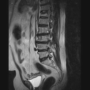
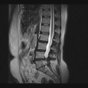
REPORT:
The imaged soft tissues show no abnormalities .
The visualized cord and filum terminalis are normal .
The conus medullaris terminates normally at L1.
Paravertebral stripe is normal in shape and signal intensity .
– Thoracolumbar scoliosis
– Decreased height of T12 with marrow signal change suggestive for recent compressive Fx
– L2/L3 spondylosis – disc dehydration , bulging & canal – foramina stenosis
– L3/L4/L5 spondylosis – disc space narrowing , dehydration , broad based protrusion & facets arthrosis – ligament flavum hypertrophy with severe canal – foramina stenosis
– L5/S1 disc dehydration & bulging with canal – foramina stenosis
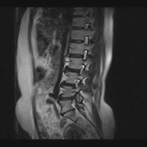
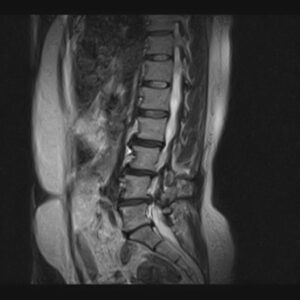
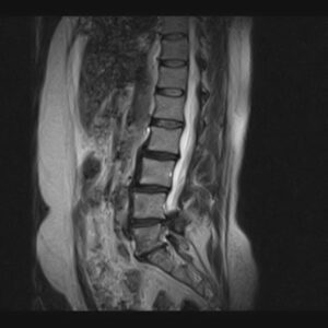
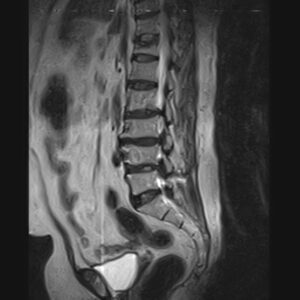
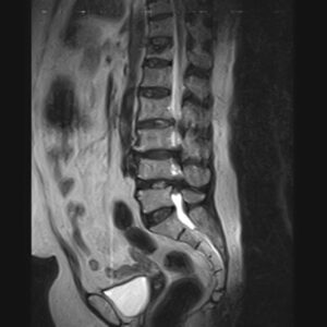
