
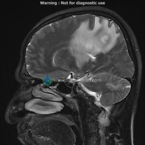
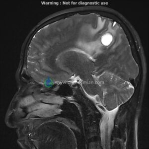
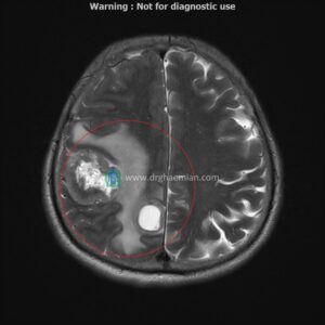
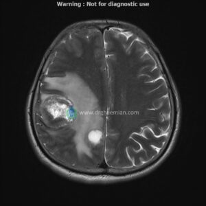
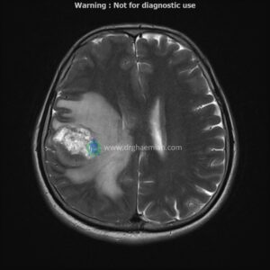
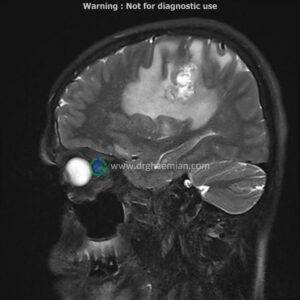
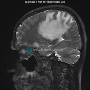
ام آر آی مغز یک روش تصویربرداری است که با استفاده از آهنربا های قوی و امواج رادیویی تصاویری از مغز و بافت های عصبی پیرامونی آن ایجاد می کند.در این کیس متاستاز مغزی و توده در مغز و… دیده میشود

گزارش پزشک
BRAIN MRI
(With & Without contrast)
Siemens MRI ( magnetom altea 1.5 tesla )
Technique : Axial T1 & T2 & FLAIR , Sagittal T2, Coronal T1; post contrast Axial fat-sat T1, Sagittal T1, Coronal T1 .
REPORT :
The interhemispheric fissure is centered on the midline .
The cerebral ventricles / cisterns are normal size and shape .
The cerebello pontine angle area appears normal on each side.
The internal acoustic meatus has normal width .
The orbital contents , falx , dura and calvaria are unremarkable .
Major intracranial vascular structures , pericavernous spaces and visualized intracranial nerve complex are normal .
The paranasal sinuses are clear and aerated with no evidence of bone erosion or destruction, fluid collection , cystic retention and mucosal thickening.
– Multiple intra-axial mass lesions (5-15mm) in both hemispheres of cerebrum & cerebellum and two large similar lesions (23mm & 43mm) in right parietal lobe with peripheral vasogenic edema & with post contrast enhancement suggestive for cerebral & cerebellar metastasis
– Pituitary gland height loss and CSF protrusion into the sella turcica consistent with partial empty sella (grade 2)
are seen.