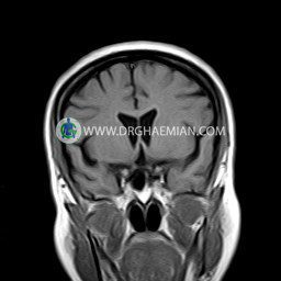
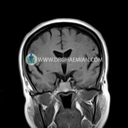
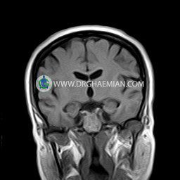
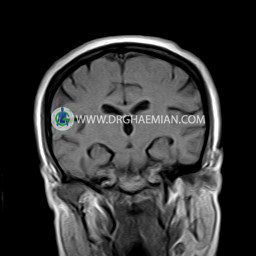
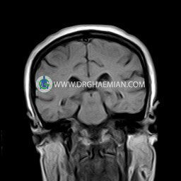
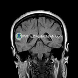
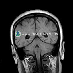
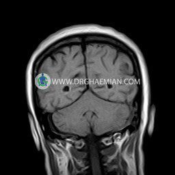
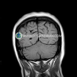
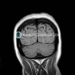
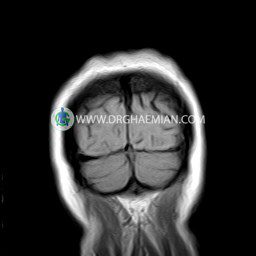
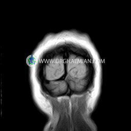
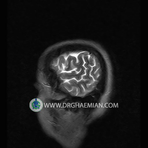
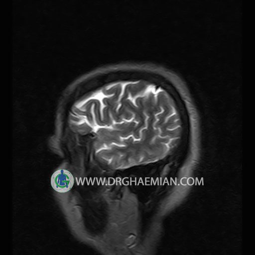
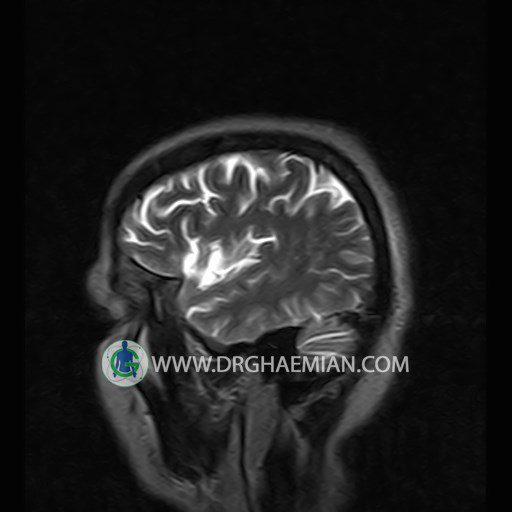
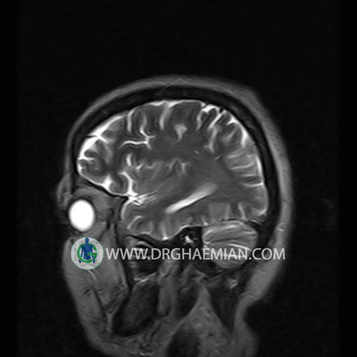
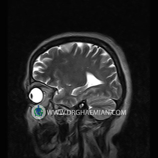
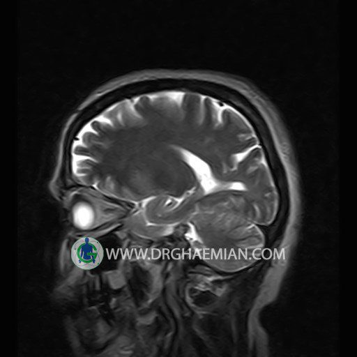
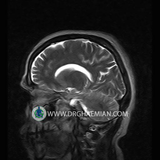
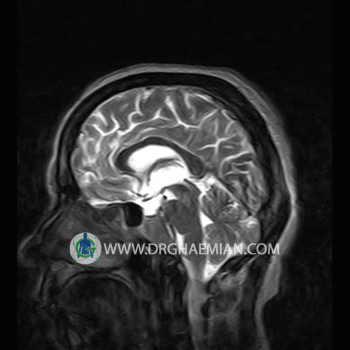
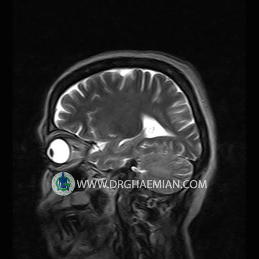
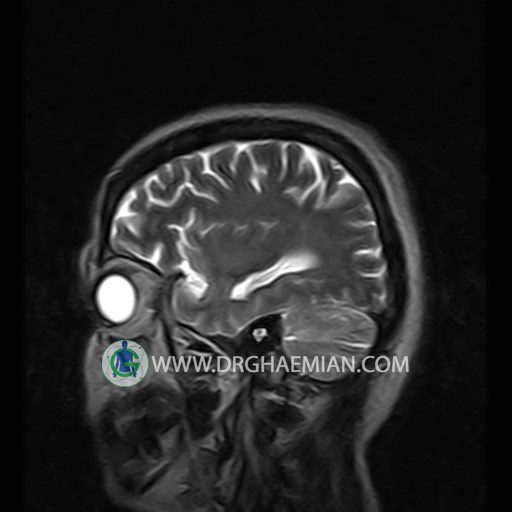
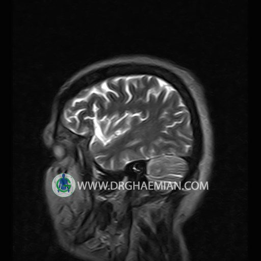
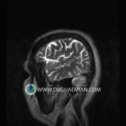
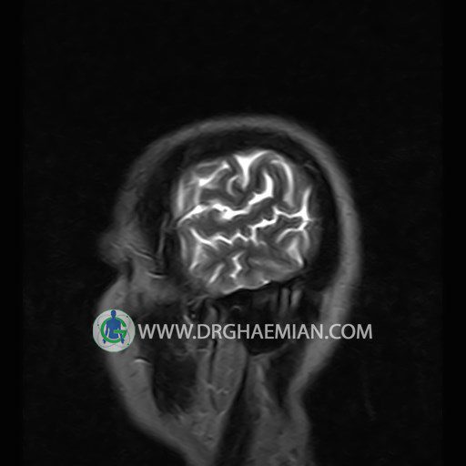
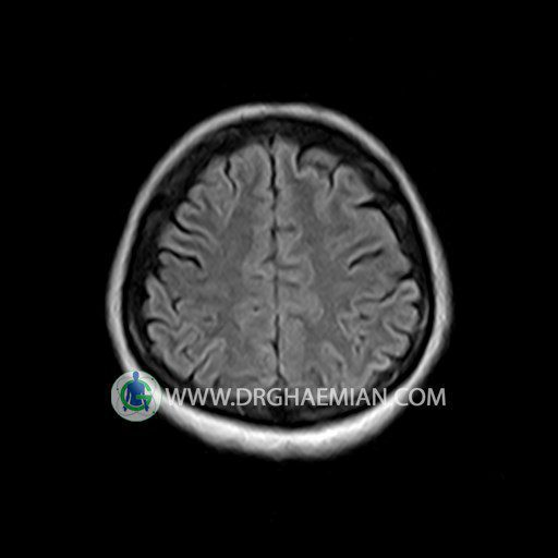
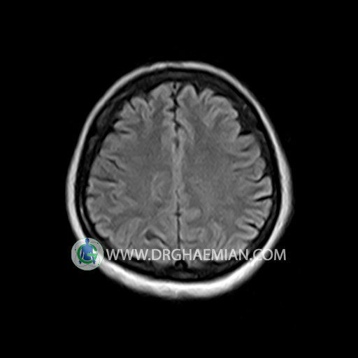
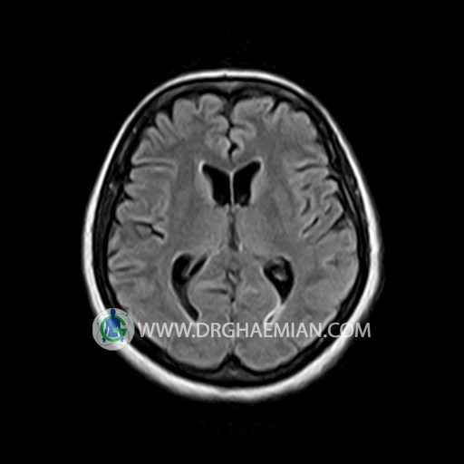
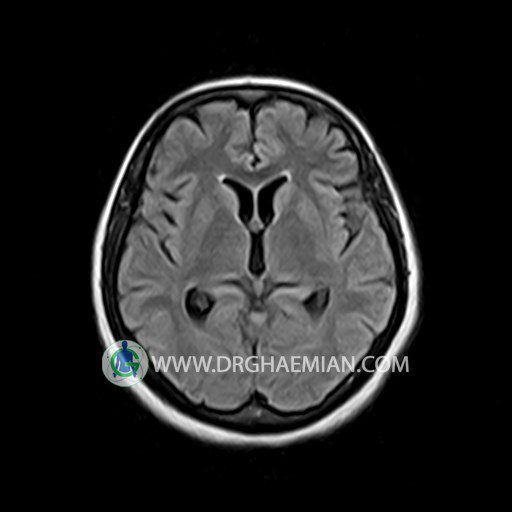
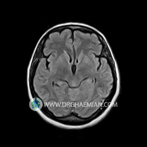
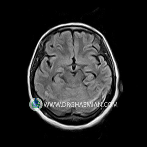
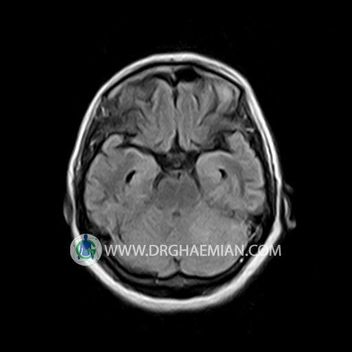
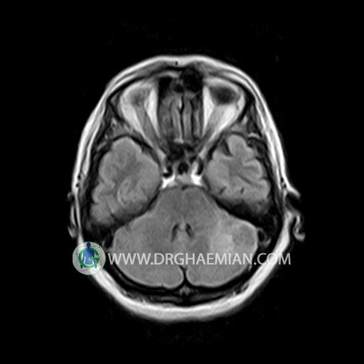
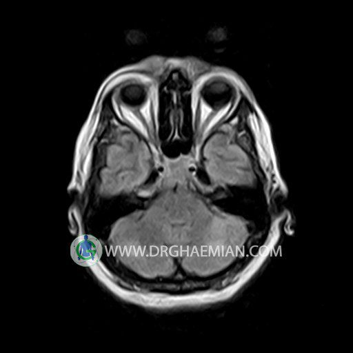
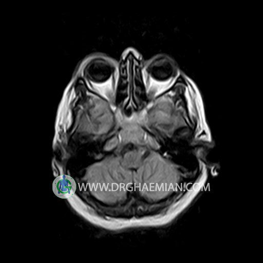
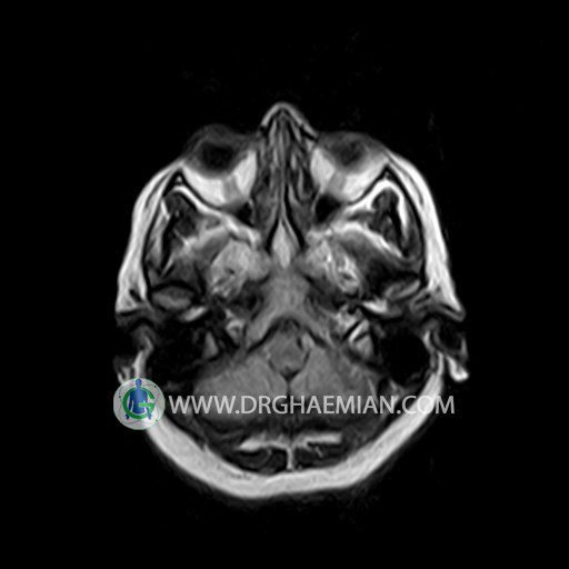
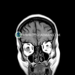
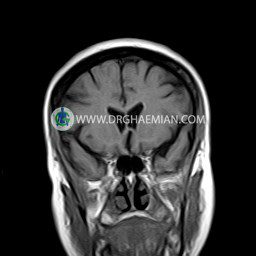
پزشکان اغلب از تصویربرداری ام آر آی برای تشخیص و درمان عارضه های پزشکی که فقط با استفاده از اشعه ایکس یا میدان مغناطیسی و امواج رادیویی قابل مشاهده است، استفاده می کنند. دستگاه ام آر آی تصاویر دقیق از ساختار های داخلی بدن ایجاد می کند. در این کیس ضایعه نئوپلاستیک مغز و سلای نسبتا خالی دیده می شود.
گزارش پزشک :
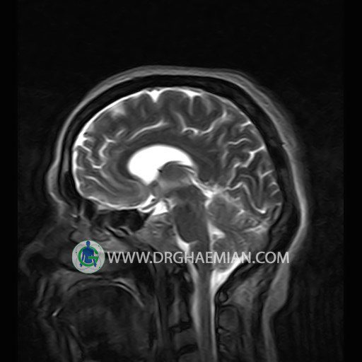
BRAIN MRI
(Without contrast)
Technique:Axial FLAIR , Axial , sagittal , FSE T2 , coronal T1 .
REPORT :
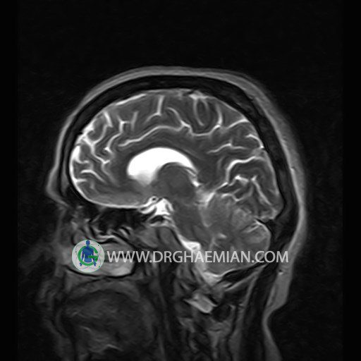
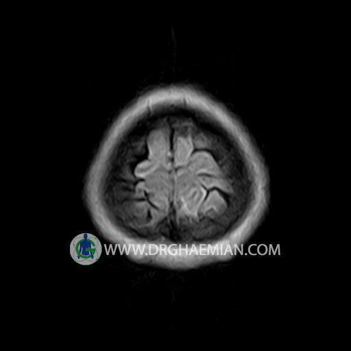
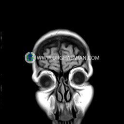
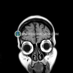
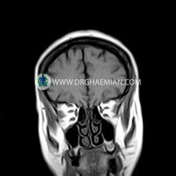
The interhemispheric fissure is centered on the midline .
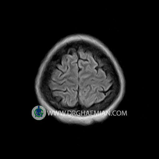
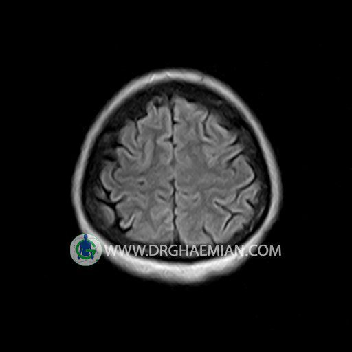
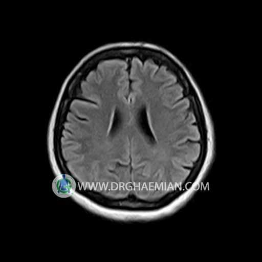
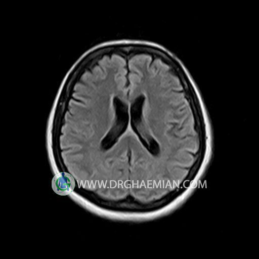
The cerebral ventricles / cisterns are normal size and shape .
No abnormalities are seen in the basal ganglia , int . capsule, corpus callosum , Thalamus and tectal plate .
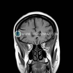
The brain stem shows no abnormal changes in signal characteristics.
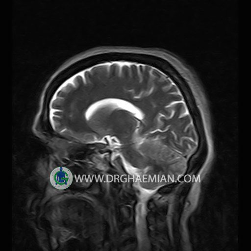
The cerebello pontine angle area appears normal on each side.
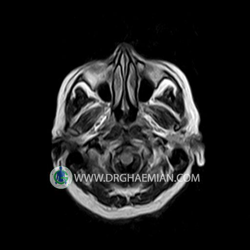
The internal acoustic meatus has normal width .
The orbital contents , falx , dura and calvaria are unremarkable .
Major intracranial vascular structures , pericavernous spaces and visualized intracranial nerve complex are normal .
The paranasal sinuses are clear and aerated with no evidence of bone erosion or destruction, fluid collection , cystic retention and mucosal thickening.
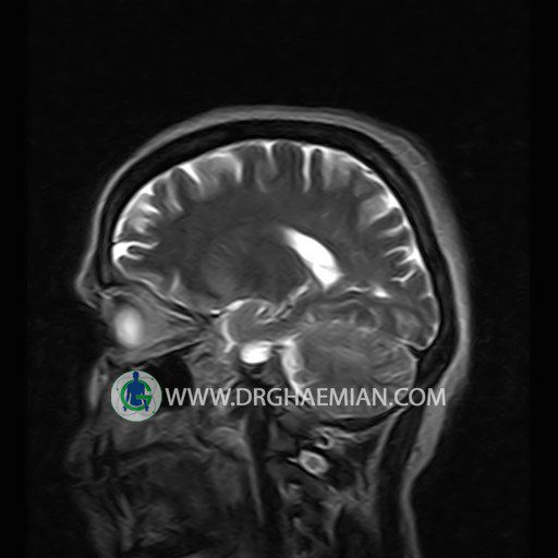
– An ill defined intra axial mass like lesion (14×15mm) in left cerebellum with mild peripheral vasogenic edema suggestive for neoplasic lesion
– Extension of suprasella cistern to sella with thin pituitary gland in floor of sella (partial empty sella)
are seen.
COMMENT: MRI with contrast is recommended.
