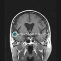
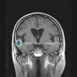
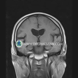
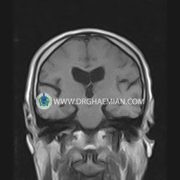
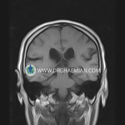
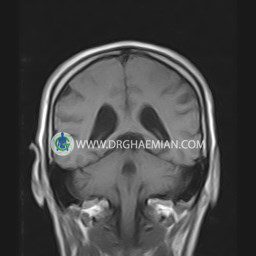
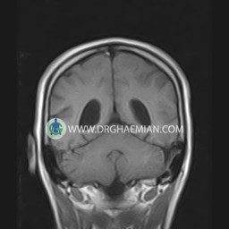
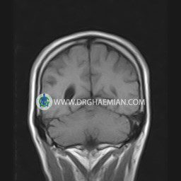
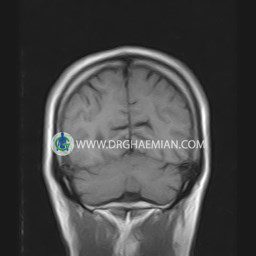
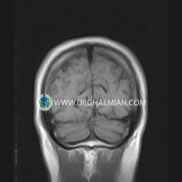
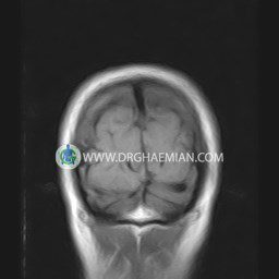
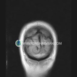
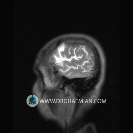
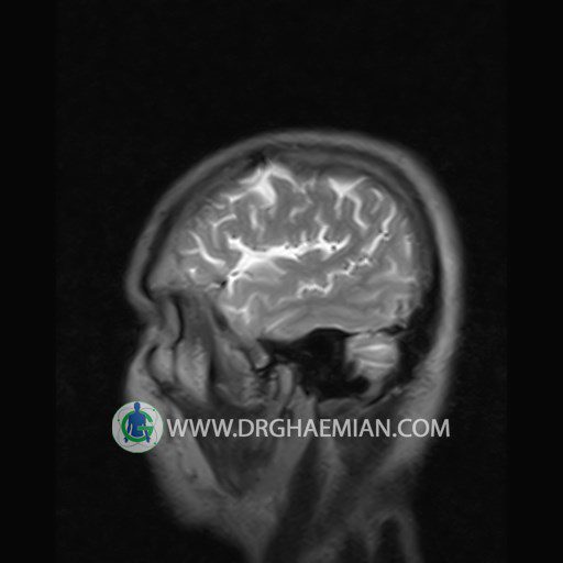
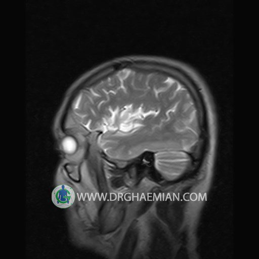
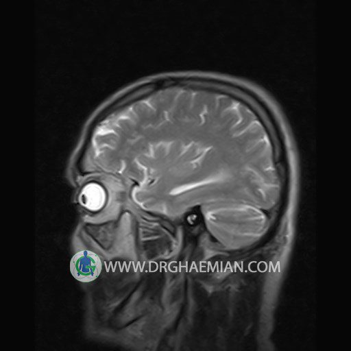
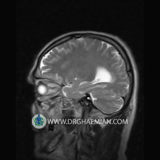
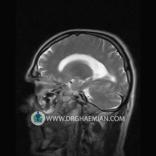
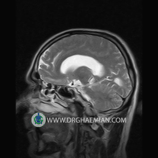
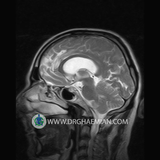
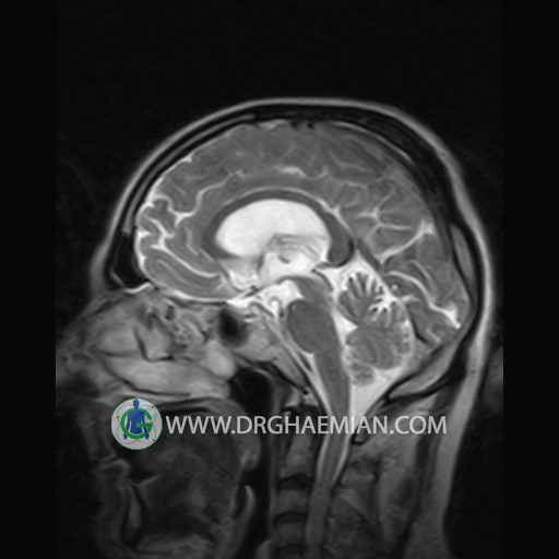
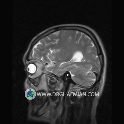
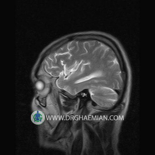
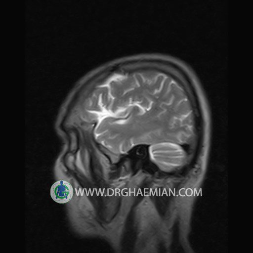
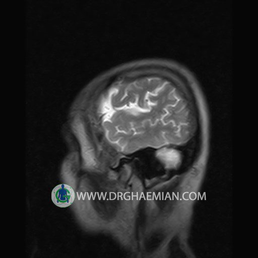
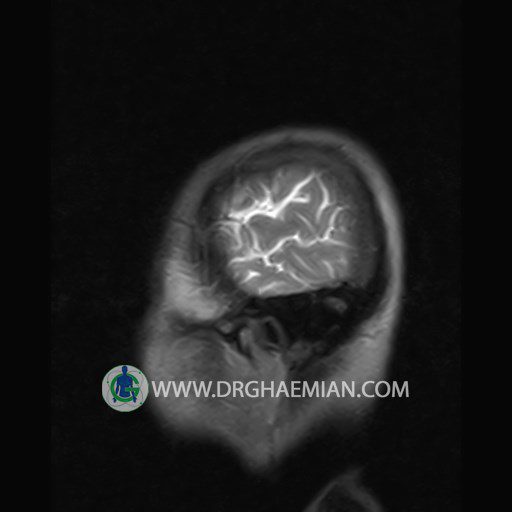
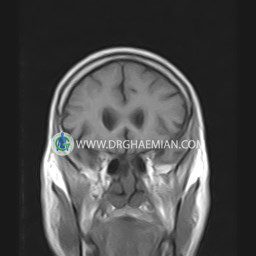
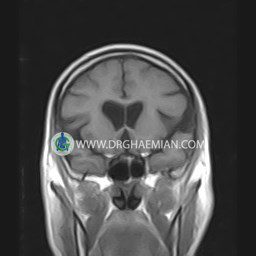
پزشکان اغلب از تصویربرداری ام آر آی برای تشخیص و درمان عارضه های پزشکی که فقط با استفاده از اشعه ایکس یا میدان مغناطیسی و امواج رادیویی قابل مشاهده است، استفاده می کنند. دستگاه ام آر آی تصاویر دقیق از ساختار های داخلی بدن ایجاد می کند. در این کیس ساختار عروقی مغز در مخچه چپ نشانه دهنده ناهنجاری های شریانی وریدی کوچک یا آنژیوم است، دیده می شود.
گزارش پزشک :
BRAIN MRI
(Without contrast)
Technique:Axial FLAIR , Axial , sagittal , FSE T2 , coronal T1 .
REPORT :
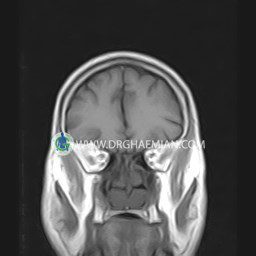
The interhemispheric fissure is centered on the midline .
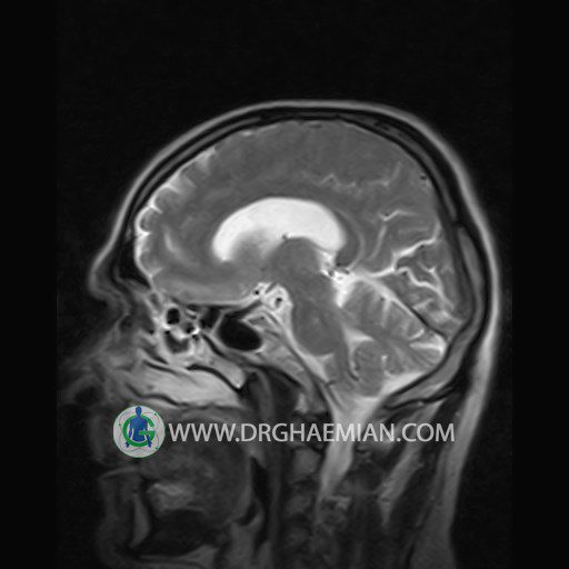
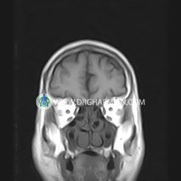
No abnormalities are seen in the basal ganglia , int . capsule, corpus callosum , Thalamus and tectal plate .
The brain stem , cerebellum show no abnormal changes in signal characteristics.
The sella and pituitary – pineal g .are normal and parasellar, suprasellar structures are unremarkable.
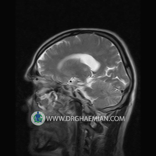
The cerebello pontine angle area appears normal on each side.
The internal acoustic meatus has normal width .
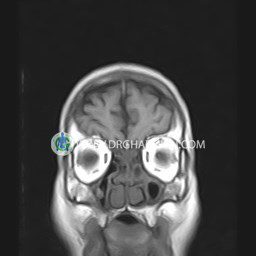
The orbital contents , falx , dura and calvaria are unremarkable .
Major intracranial vascular structures , pericavernous spaces and visualized intracranial nerve complex are normal .
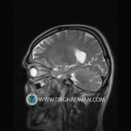
The paranasal sinuses are clear and aerated with no evidence of bone erosion or destruction, fluid collection , cystic retention and mucosal thickening.
– Heterogeneous signal change in left cerebellum suggestive for vascular structure such as small AVM & angioma
is seen
COMMENT: MRI with contrast & CT angiography is recommended .
