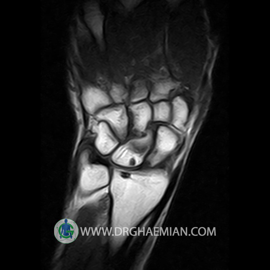
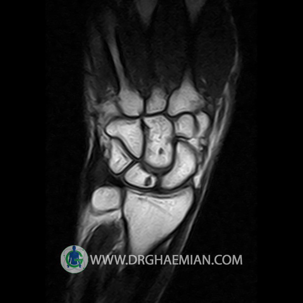
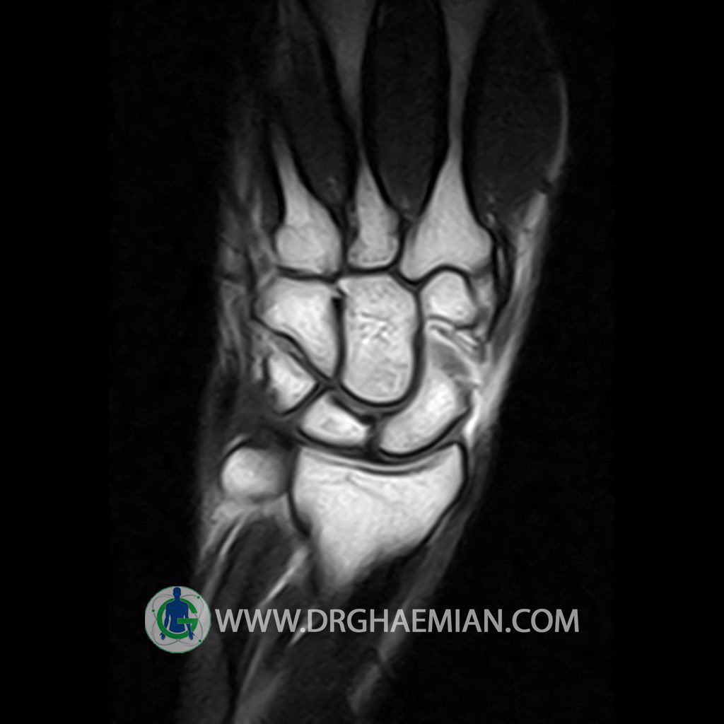
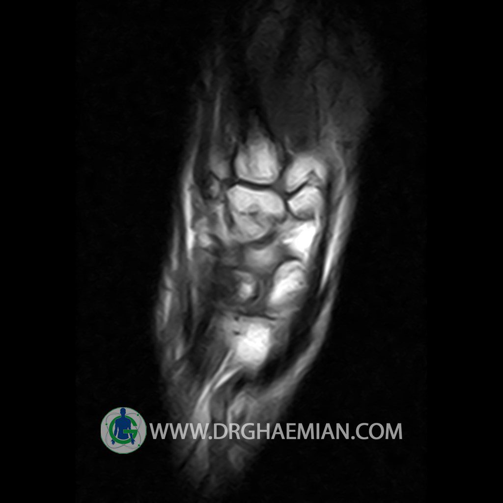
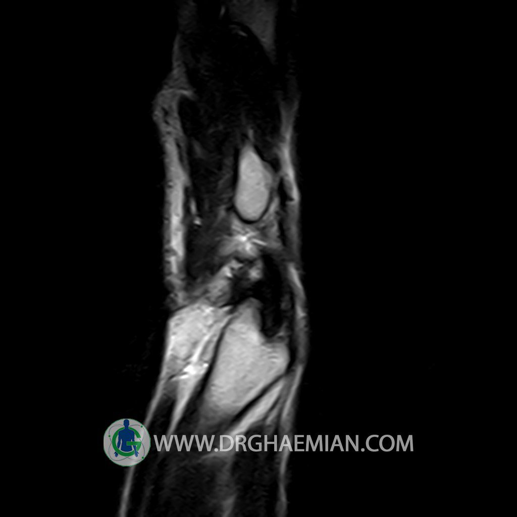
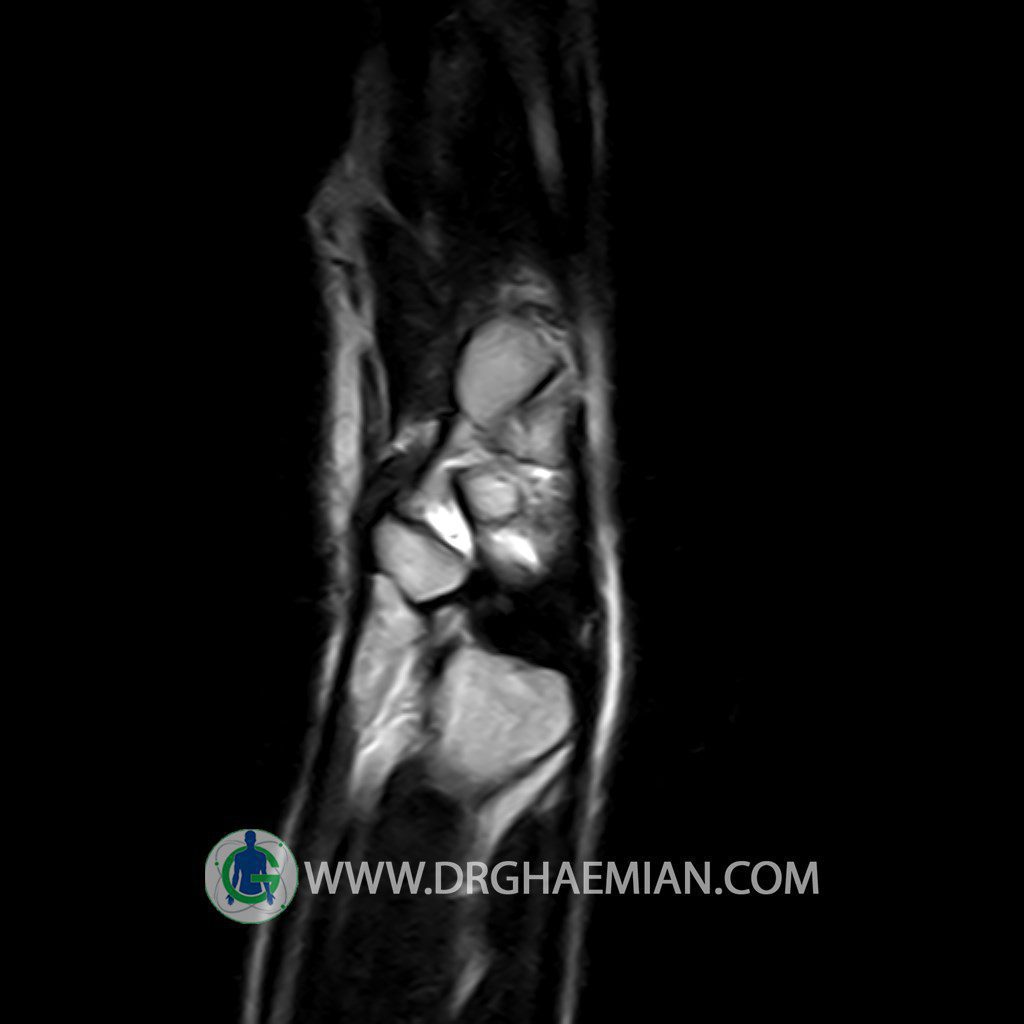
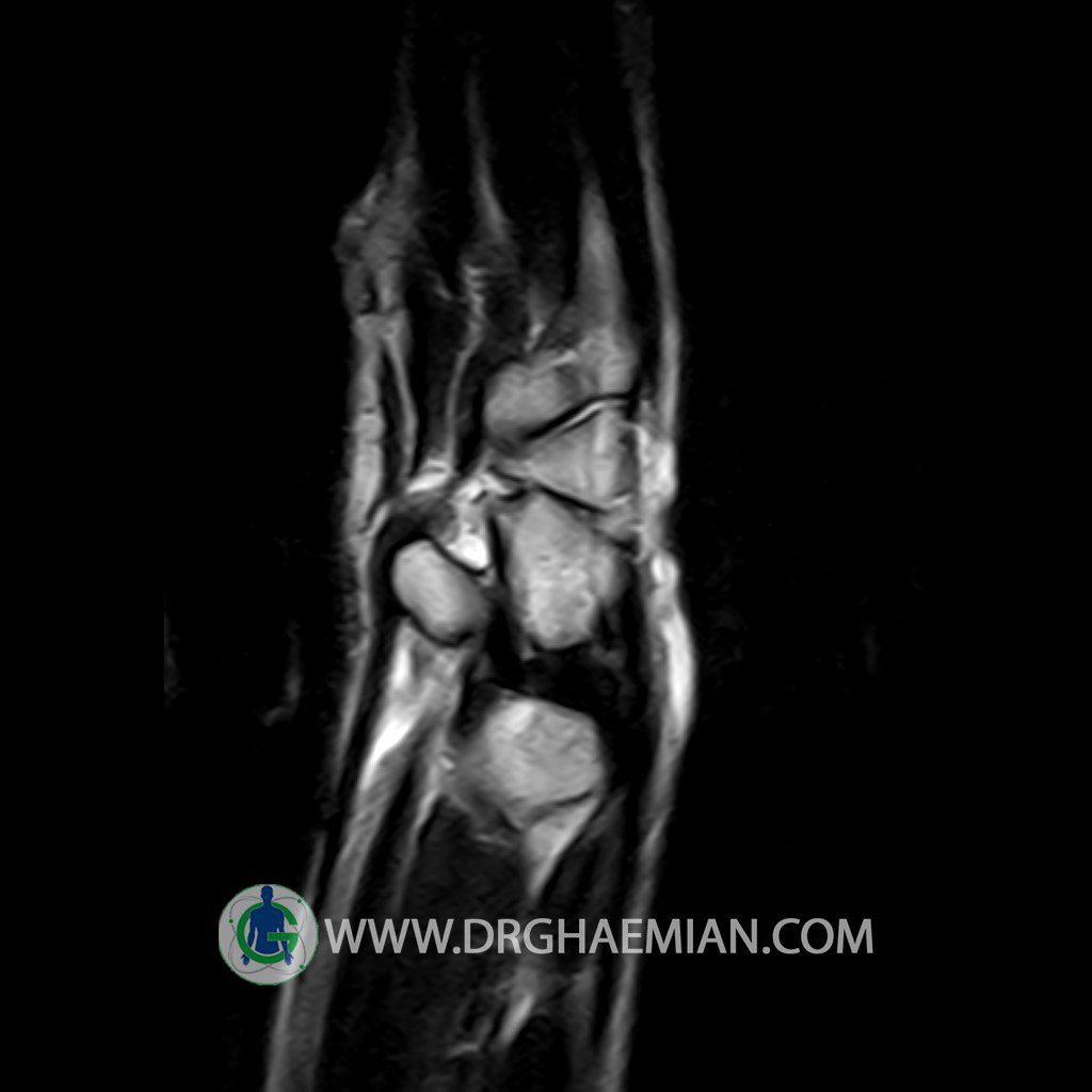
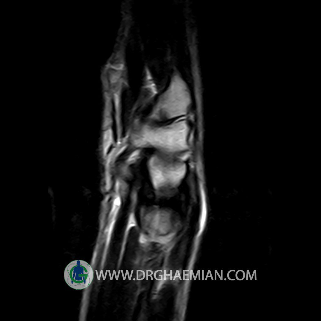
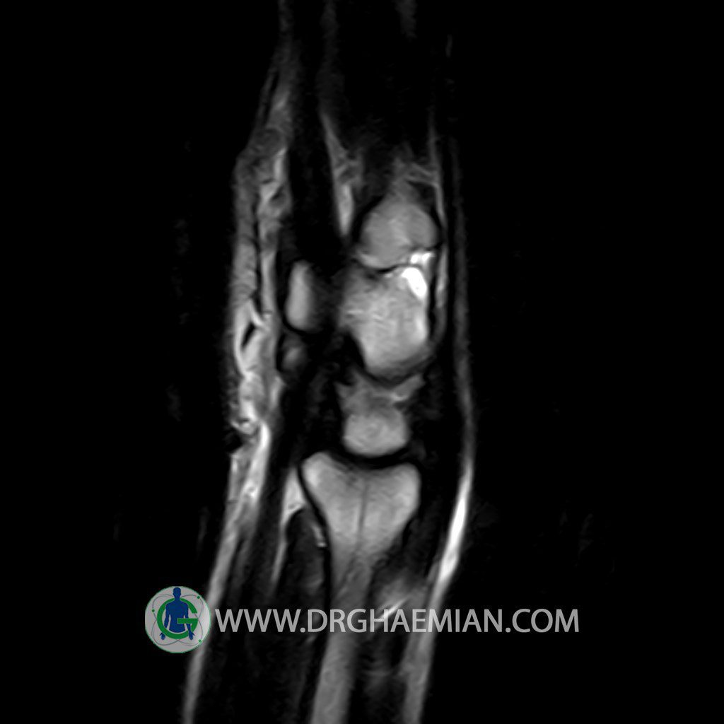
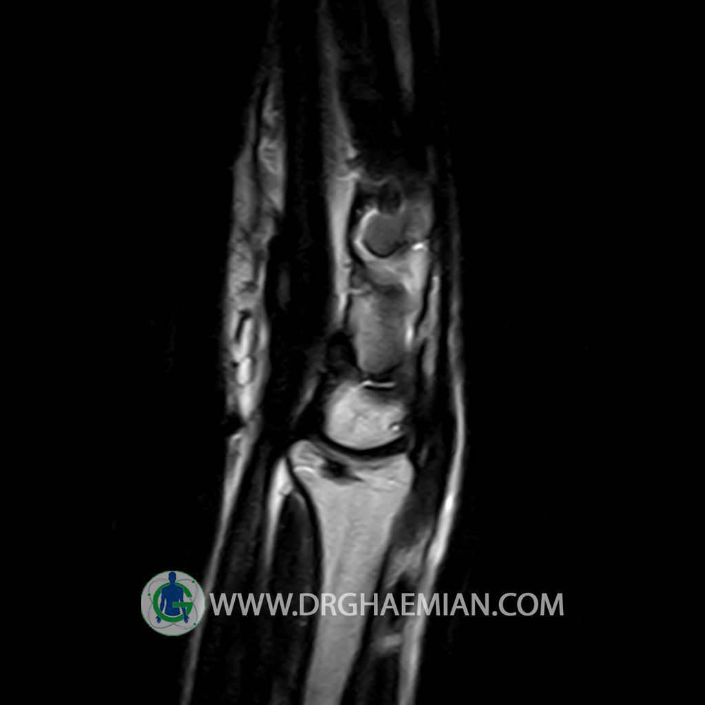
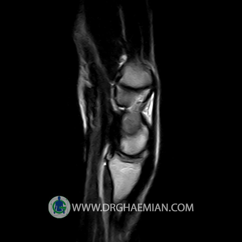
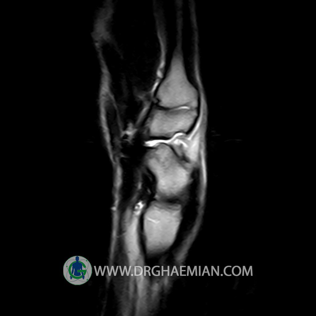
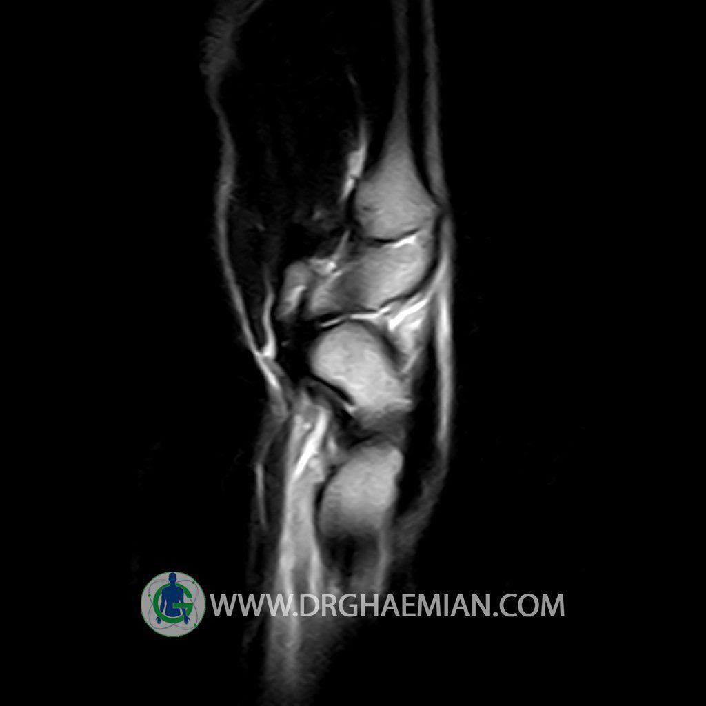
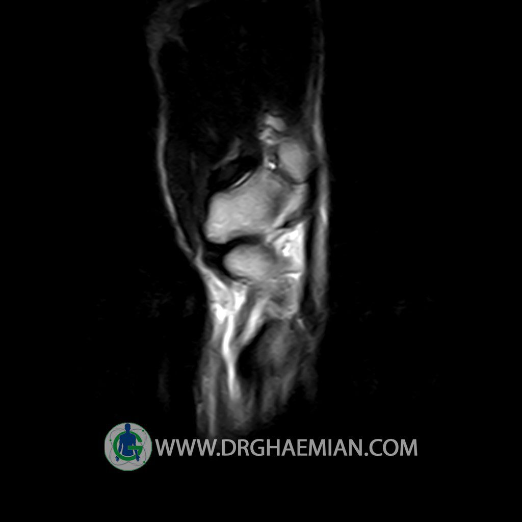
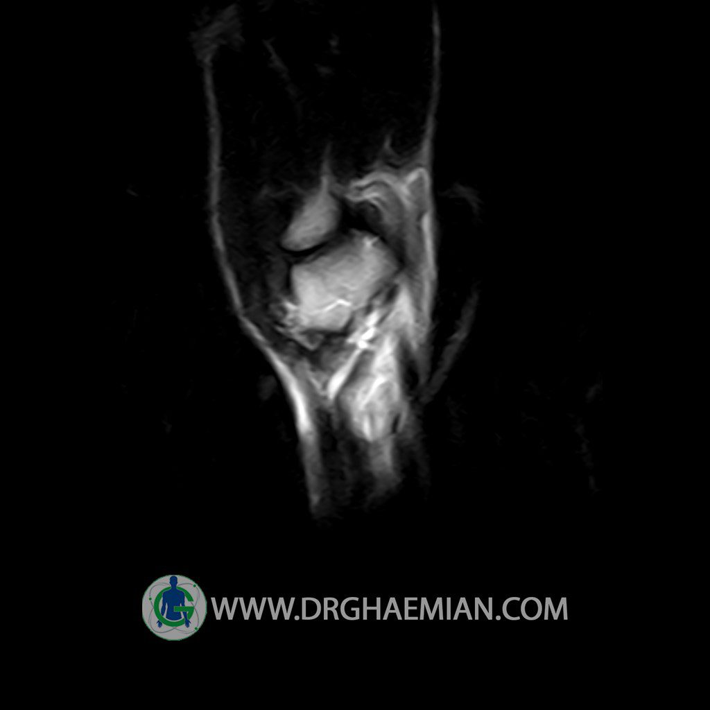
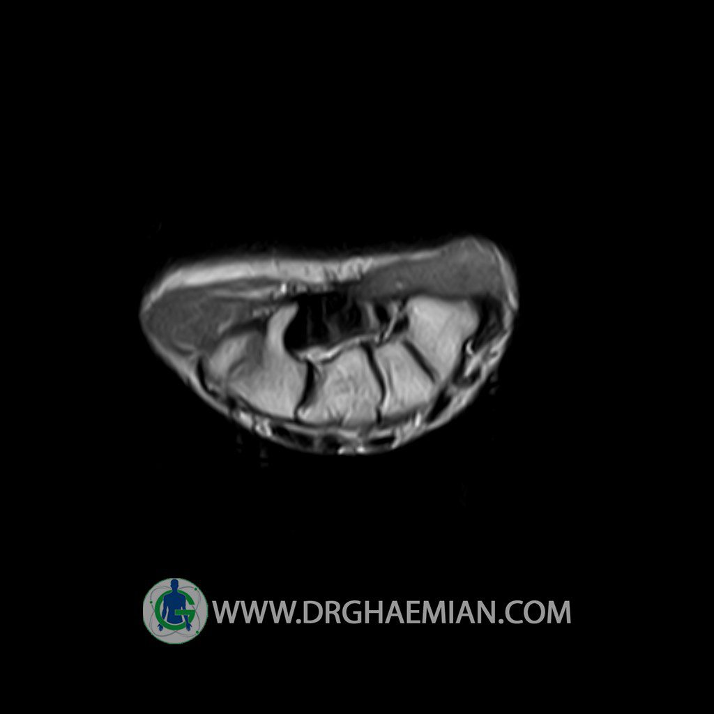
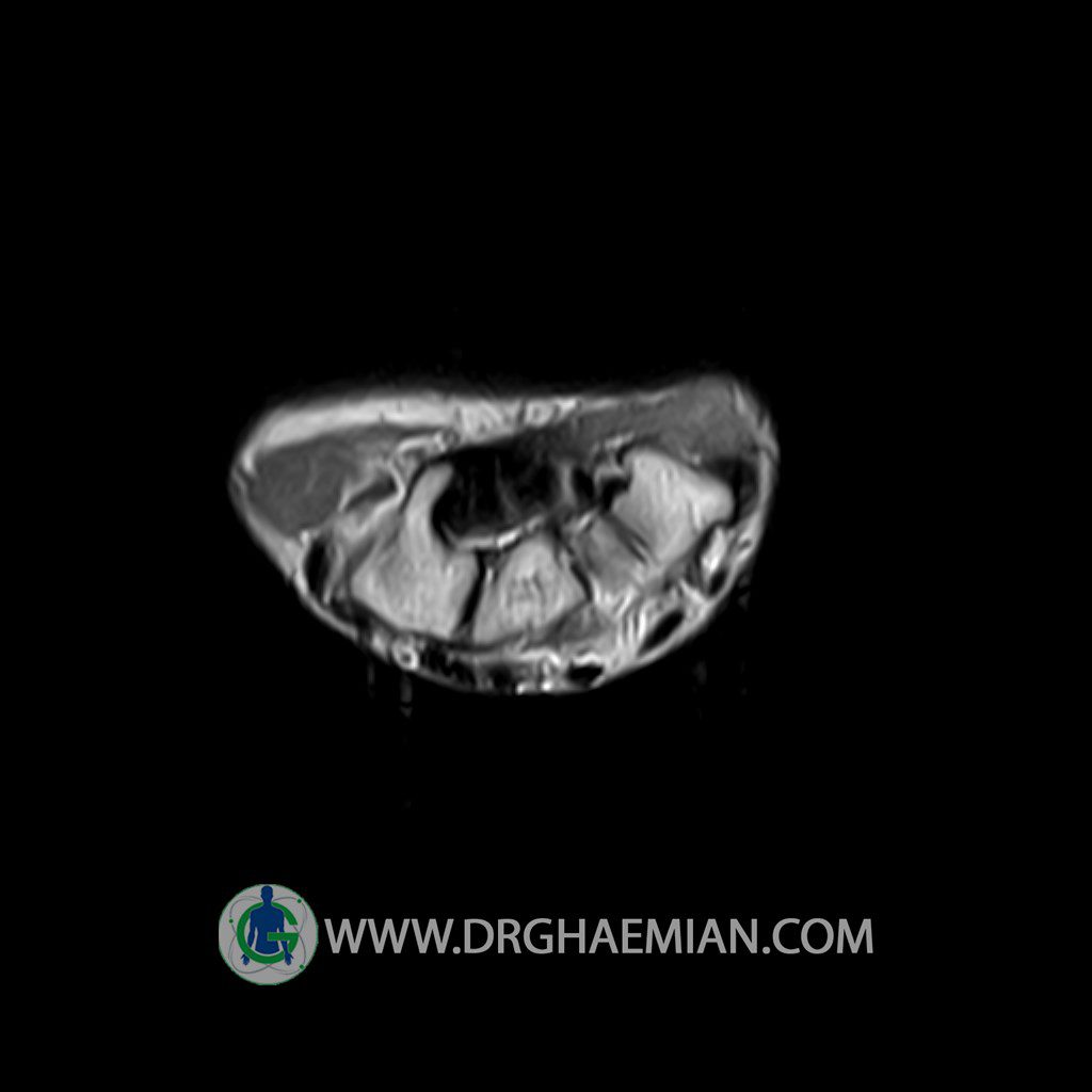
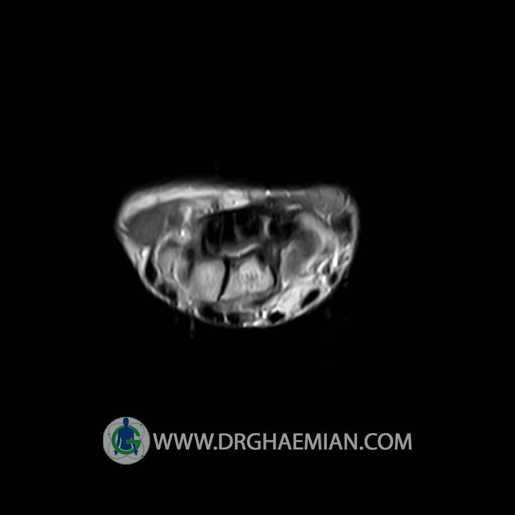
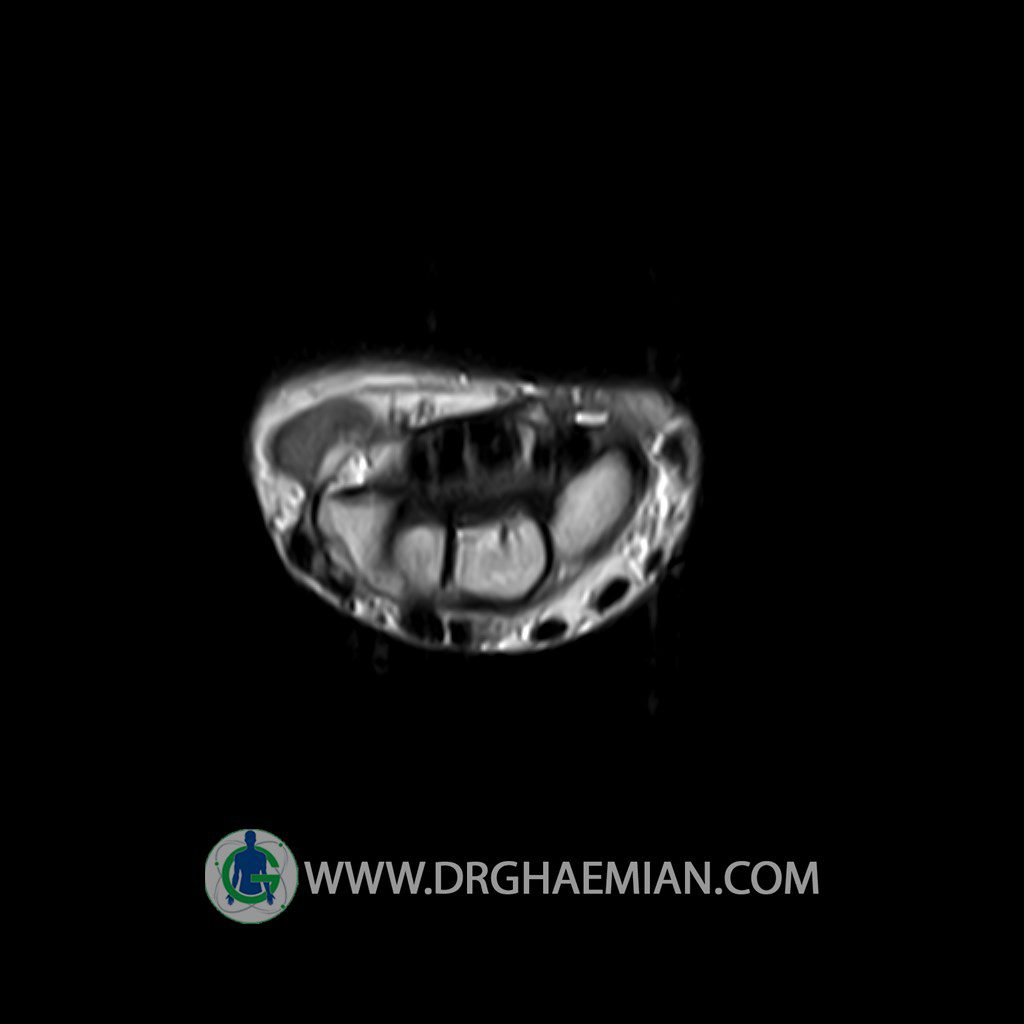
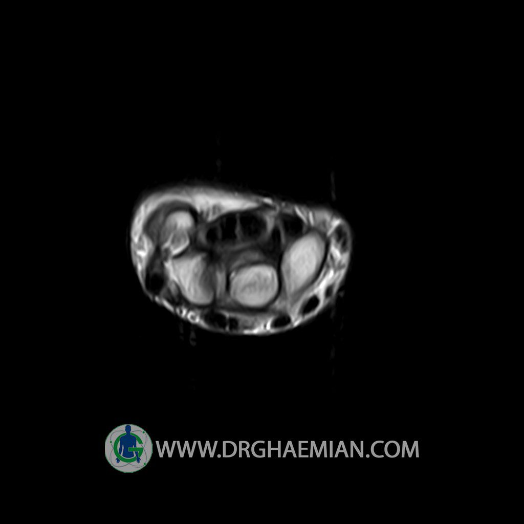
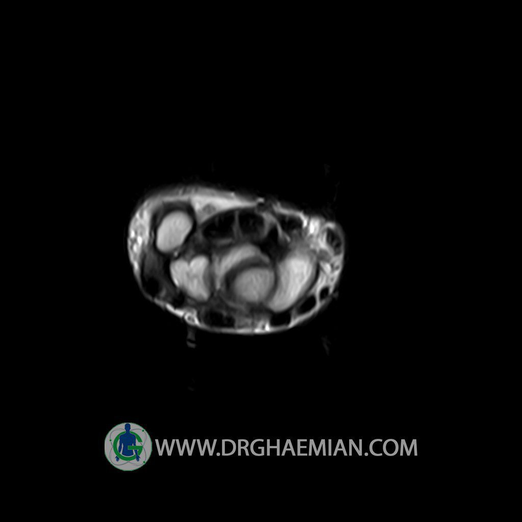
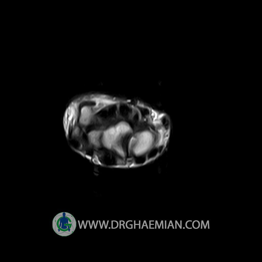
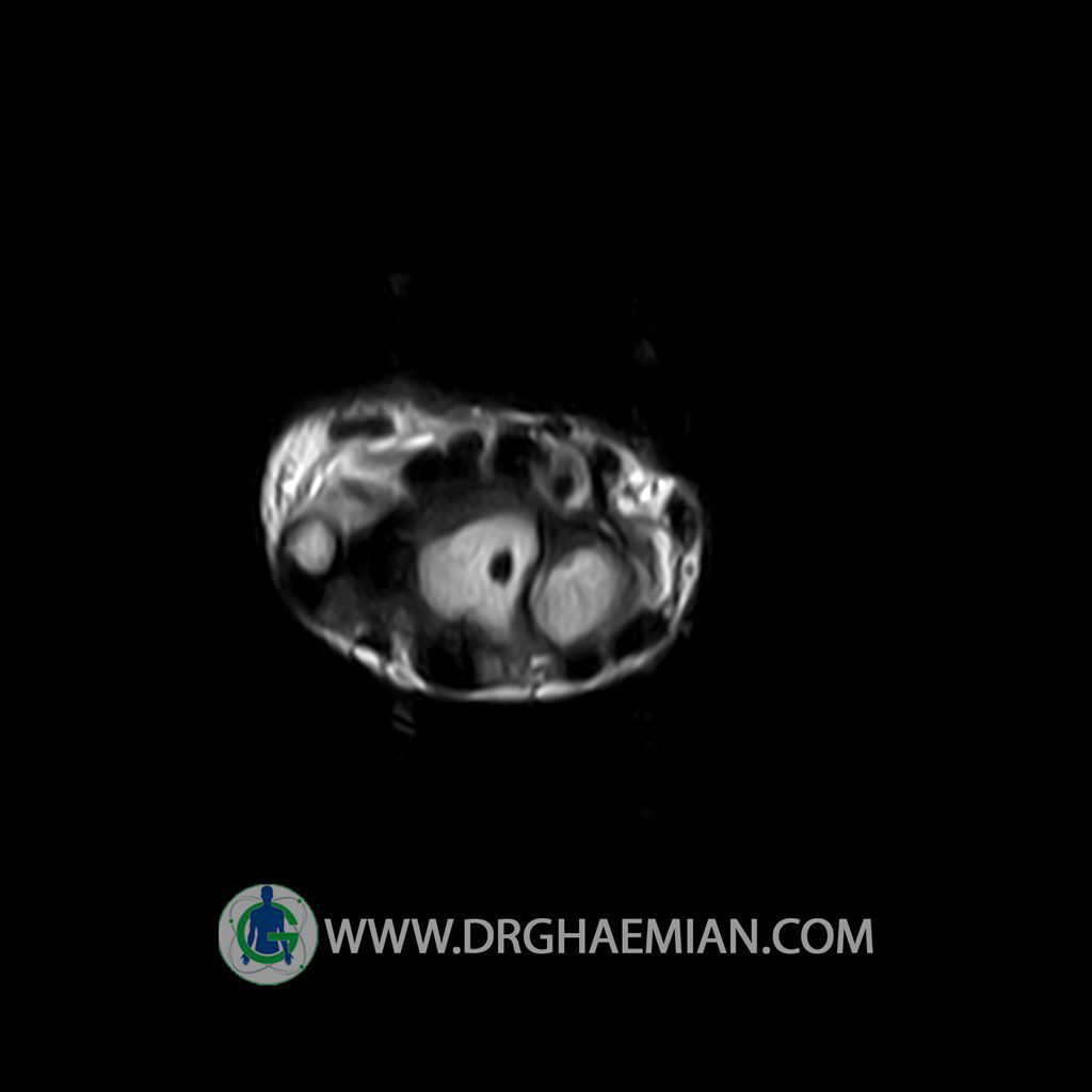
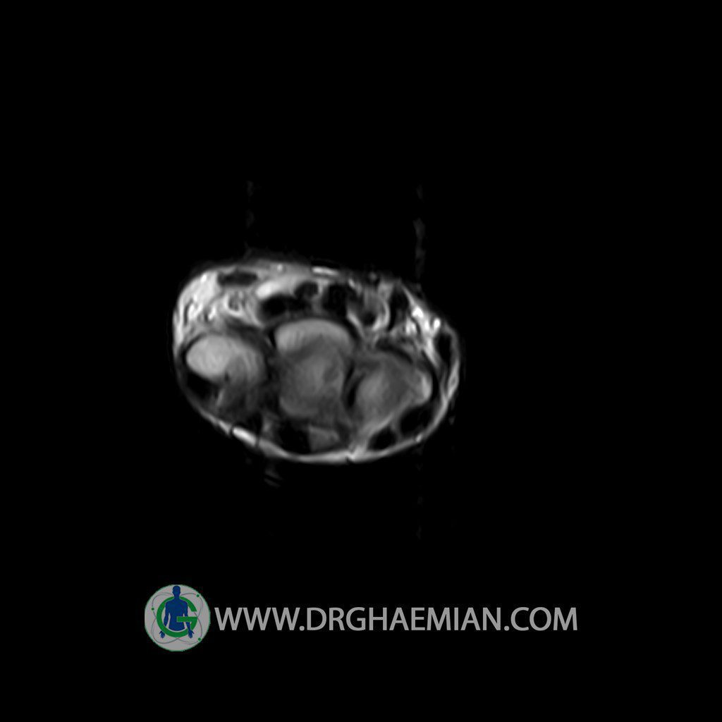
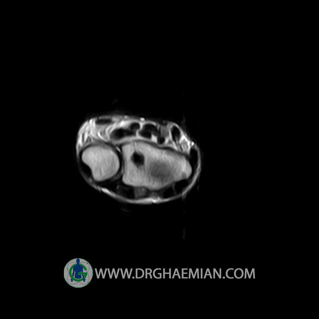
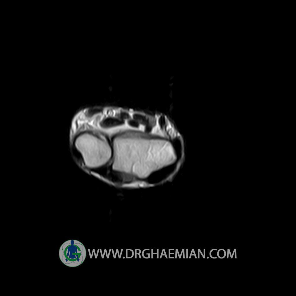
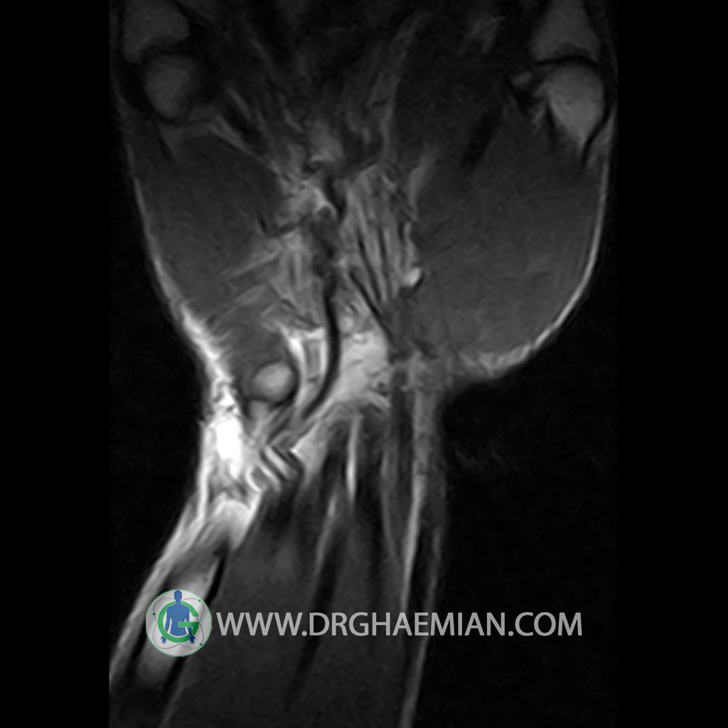
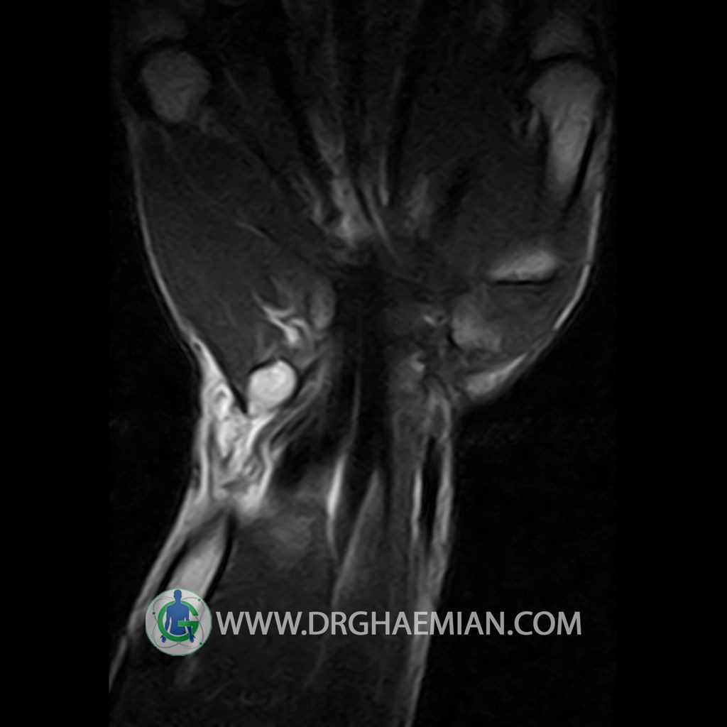
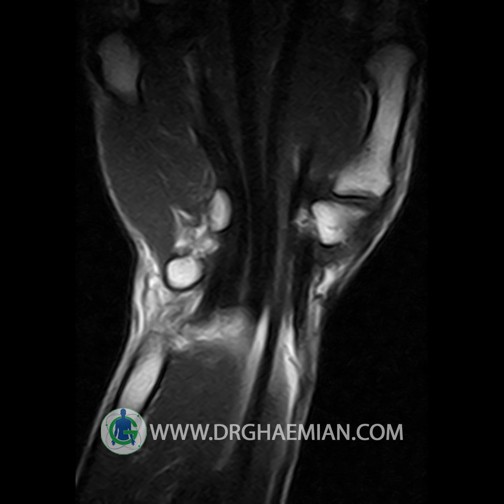
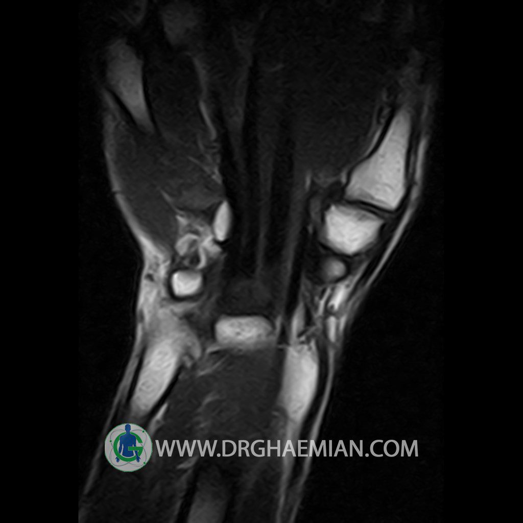
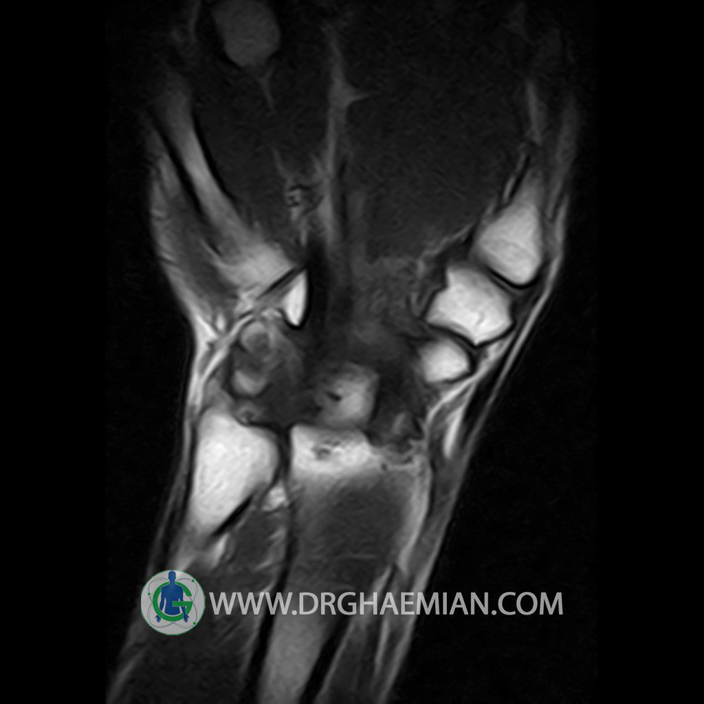
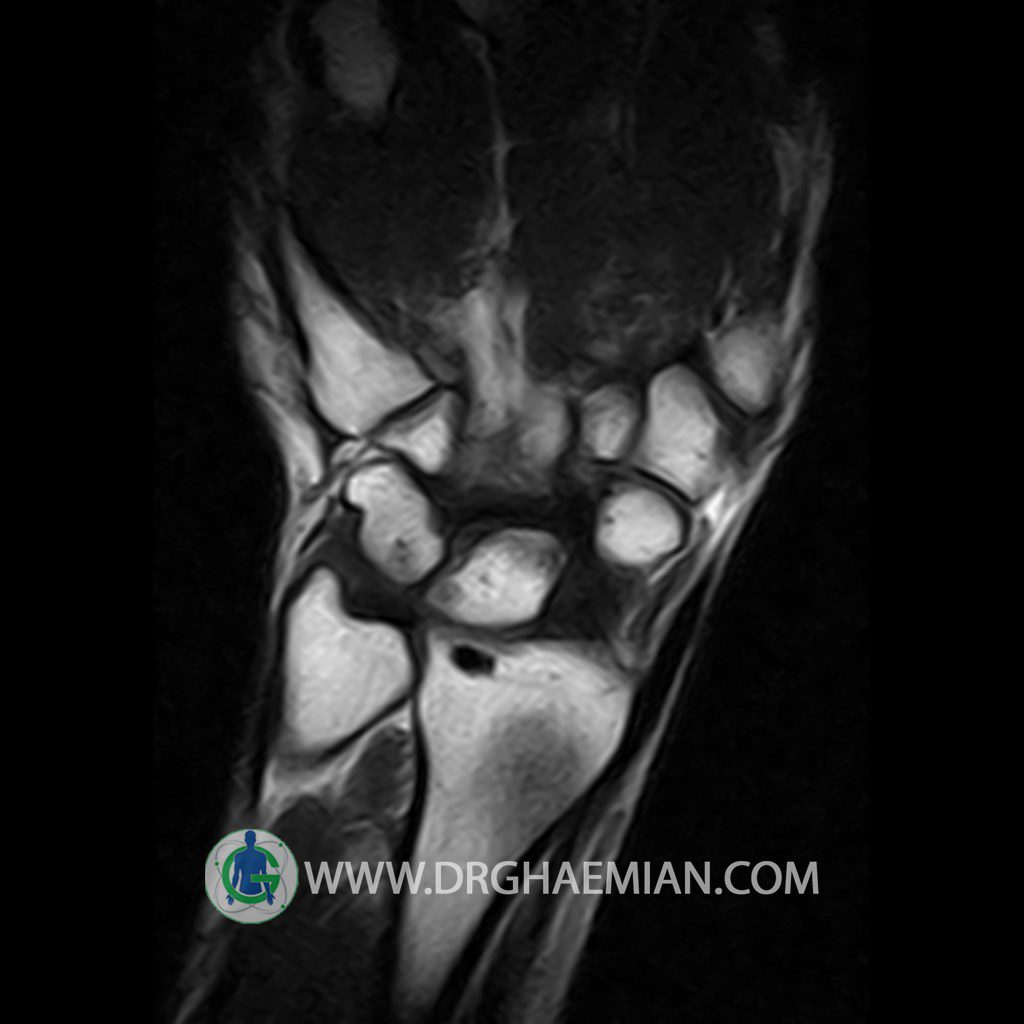
پزشکان اغلب از تصویربرداری ام آر آی برای تشخیص و درمان عارضه های پزشکی که فقط با استفاده از اشعه ایکس یا میدان مغناطیسی و امواج رادیویی قابل مشاهده است، استفاده می کنند. دستگاه ام آر آی تصاویر دقیق از ساختار های داخلی بدن ایجاد می کند. در این کیس ضایعه اسکلروتیک مچ دست و bone island دیده می شود.


گزارش پزشک:
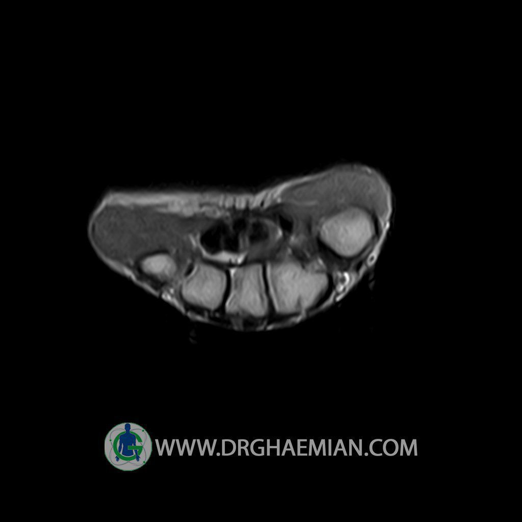
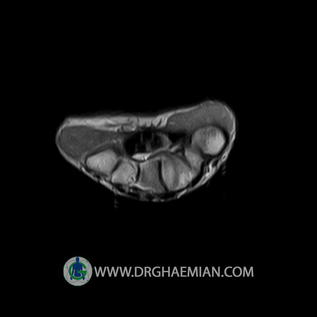
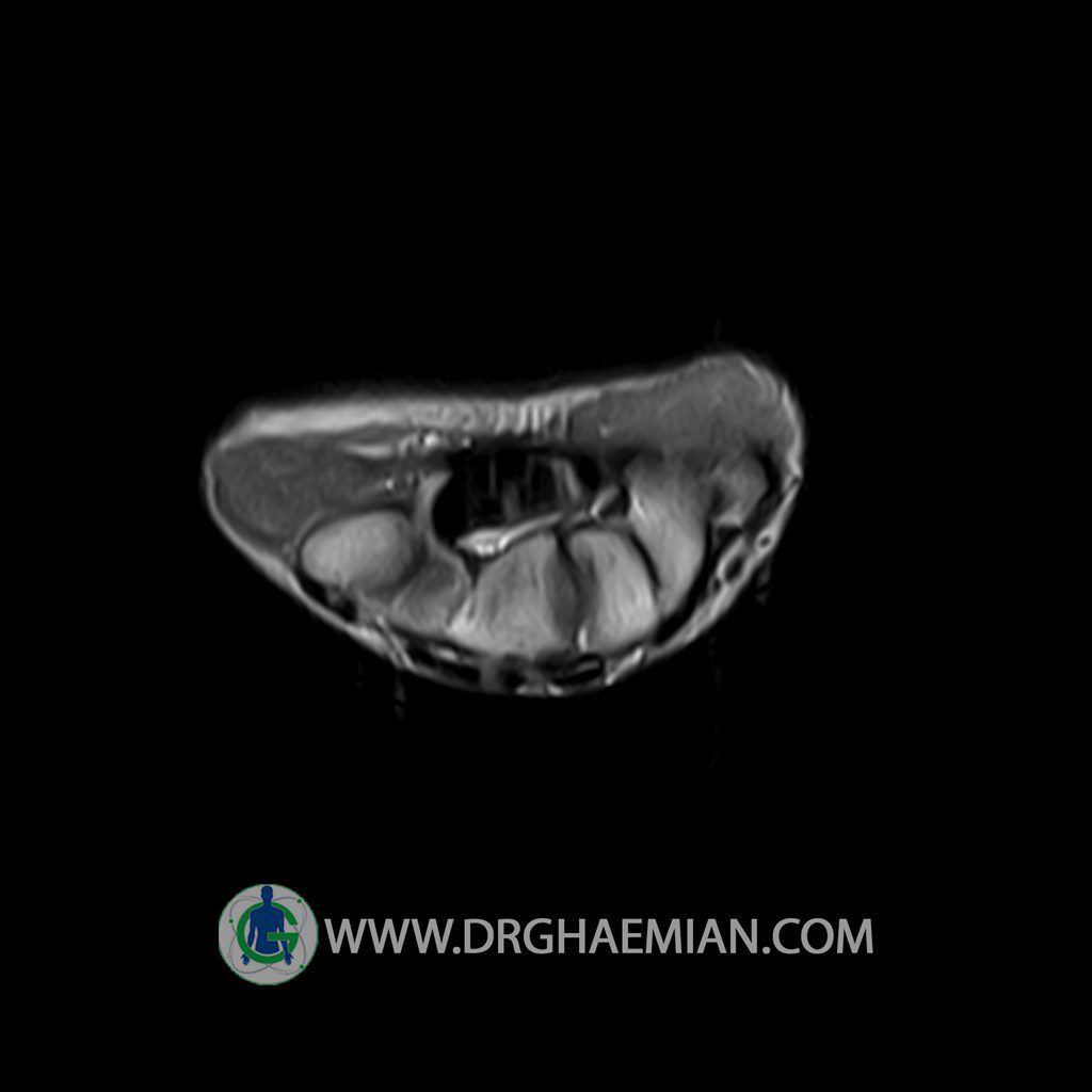
RIGHT WRIST MRI
(Without contrast)
Technique :Axial , sagittal T1 and T2 , coronal T1 , coronal SPGR .
REPORT:
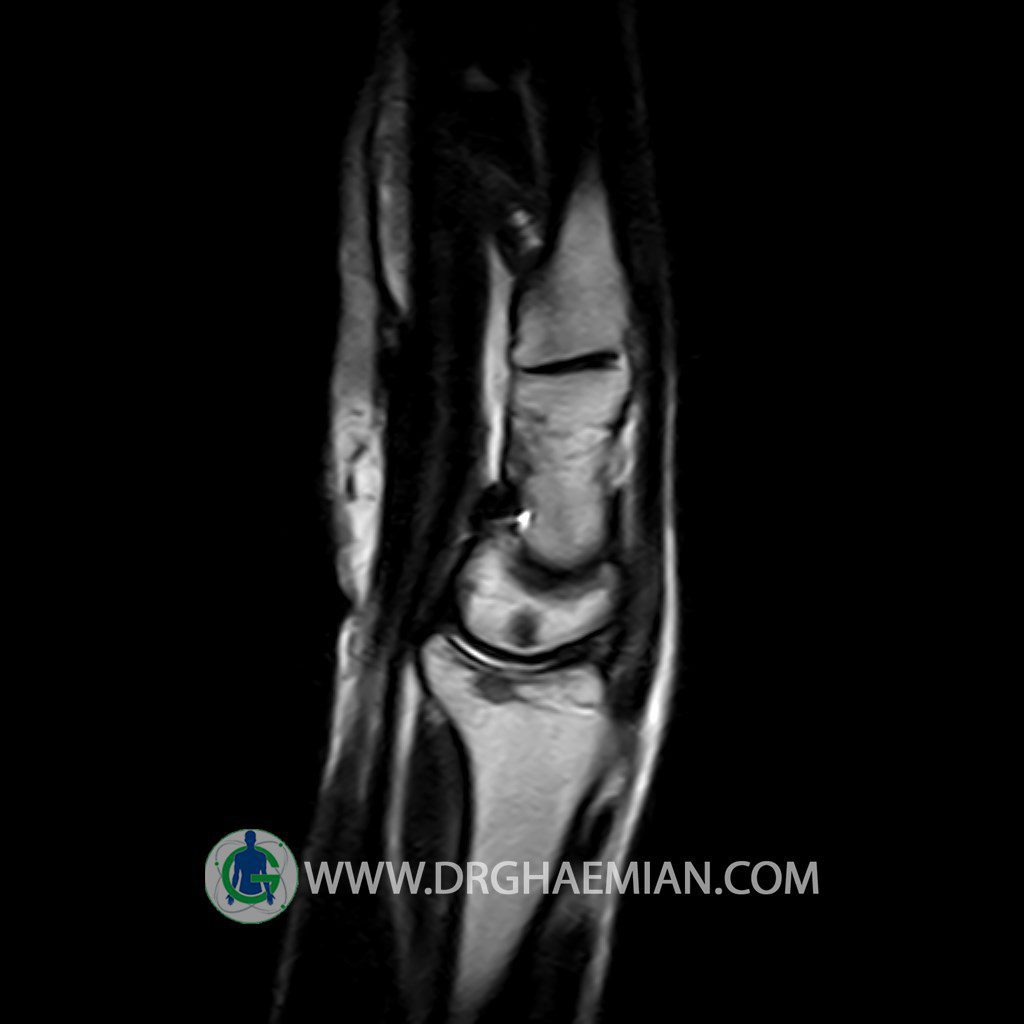
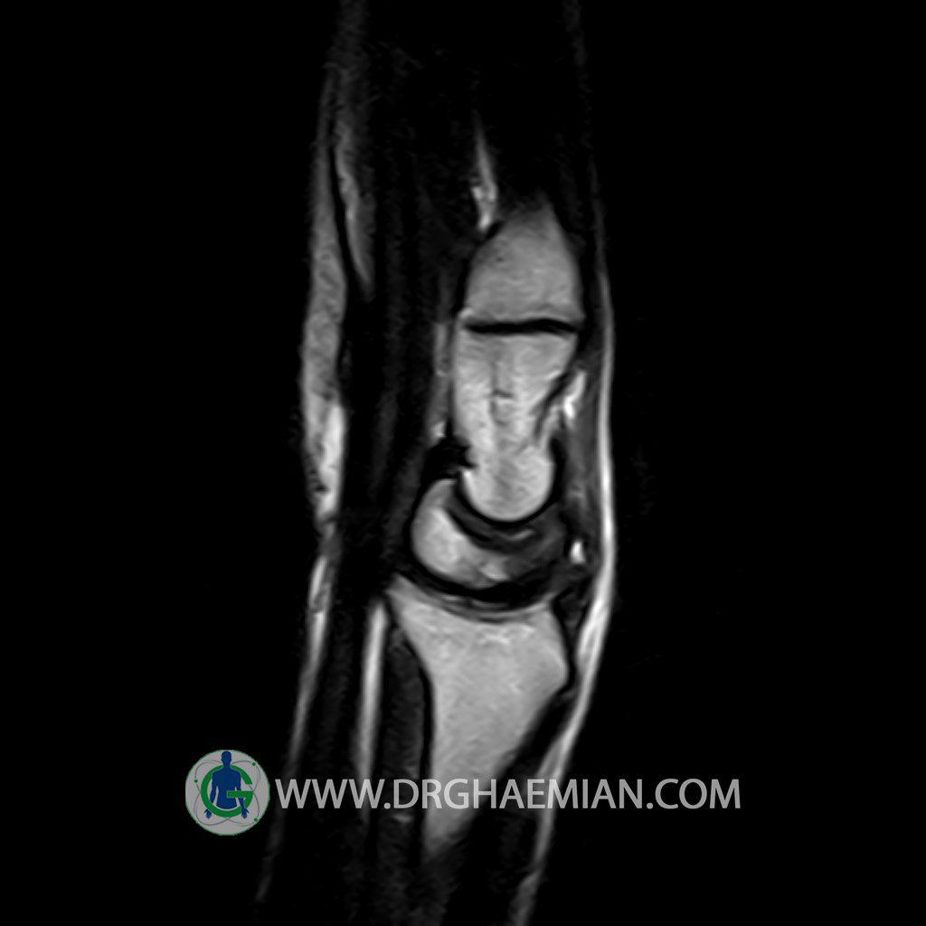
The bones comprising the wrist present a normal configuration .
The radial joint angle is normal .
The carpal bones show normal shape and relationship to one another and to the radiocarpal and carpometacarpal joints .
The articular surfaces are smooth and congruent with normal cortical thickness and normal width of joint space .
The ulnar ( triangular ) disk exhibits normal configuration and normal signal characteristics .
The interosseous ligaments also appear normal .
The carpal tunnel is normal width and transmits tendons that are normal in width and position.
Ulnar variance is natural .
– Focal signal change in distal epiphysis of radius and in lunate suggestive for sclerotic lesion and bone island
– Mild soft tissue around the wrist
are seen.
