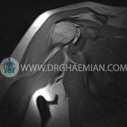
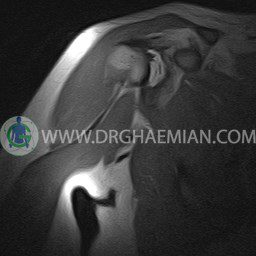
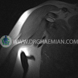
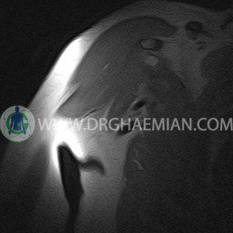
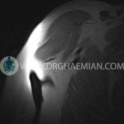
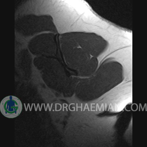
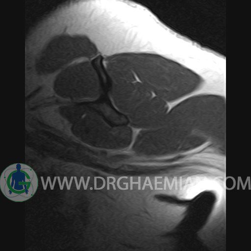
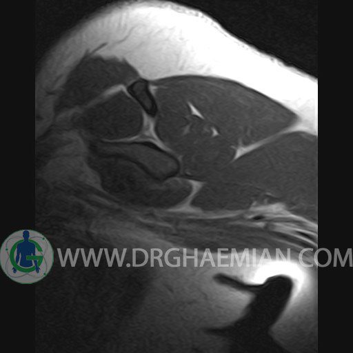
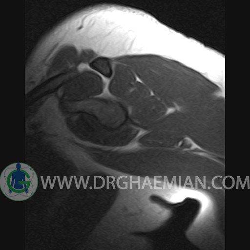
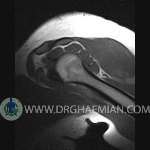
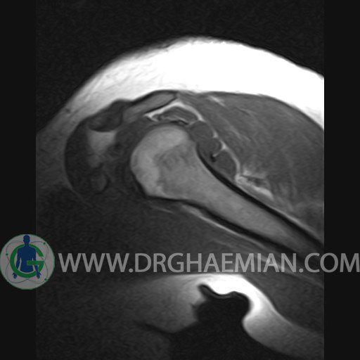
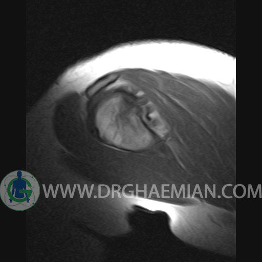
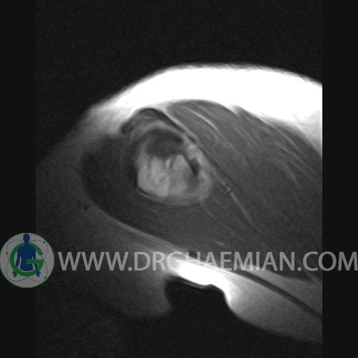
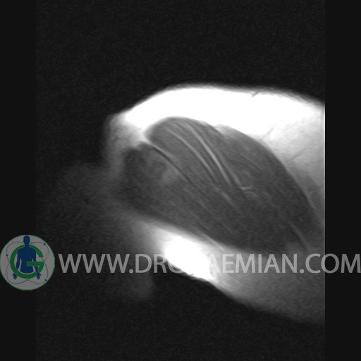
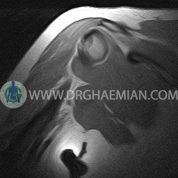
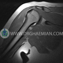
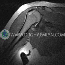
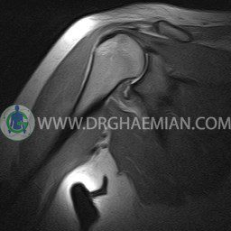
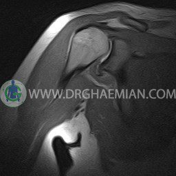
ام ار آی شانه یک روش تصویربرداری است که به وسیله آهنرباهای قدرتمند از قسمت شانه تصاویری ایجاد می کند. این نوع تصویربرداری از تششعات استفاده نمی کند. در این کیس بدشکلی هیل ساکس، سینوویت گذرا و جراحت نرم نوع بانک مشاهده می شود.


گزارش پزشک :
RIGHT SHOULDER MRI
(Without contrast)
Technique: Axial T1 and GE , coronal T1 , T2 , sagittal T1 , T2 .
REPORT:
The articular surfaces are smooth and show normal cortical thickness .
The width of the joint space is normal .
The glenoid labrum is intact on all sides .
The acromioclavicular joint has normal configuration , with no hypertrophy .
The subacromial fat is intact .
The muscles comprising the rotator cuff show normal course and configuration .
In particular , the supraspinatus muscle is normal in its position , width , and signal characteristics and shows a normal musculotendinous junction .
The biceps tendon appears normal and occupies a normal position in the bicipital groove.
The other muscles that cover the shoulder joint appear normal , as do imaged portions of the lungs and soft tissues .
– Glenohumeral joint effusion suggestive for transient synovitis
– Focal bone bruise with cortical depression at posterolateral of humeral head suggestive for hill sachs deformity
– Focal signal change in anteroinferior of labrum suggestive for soft bankart injury
are seen
