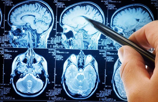
پر بازدیدترین مقالات
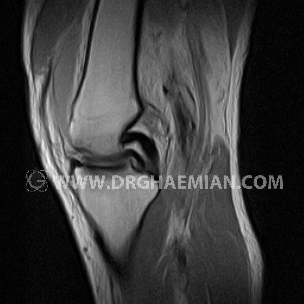
ام آر آی از پارگی رباط زانو بیمار
0
ام آر آی از زانو با استفاده از آهنرباهای قوی تصاویری از مفصل، عضلات و بافت های زانو ایجاد می…
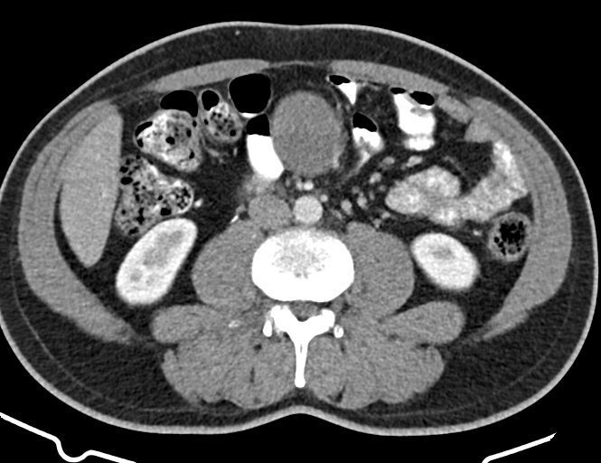
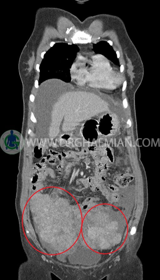
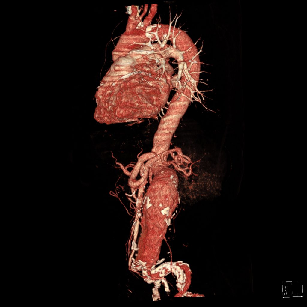
سی تی اسکن کیست مزانتریک
سی تی اسکن کیست مزانتریک در مواقعی مورد استفاده است که پزشک به مشکلی در ناحیه شکم مشکوک است اما…
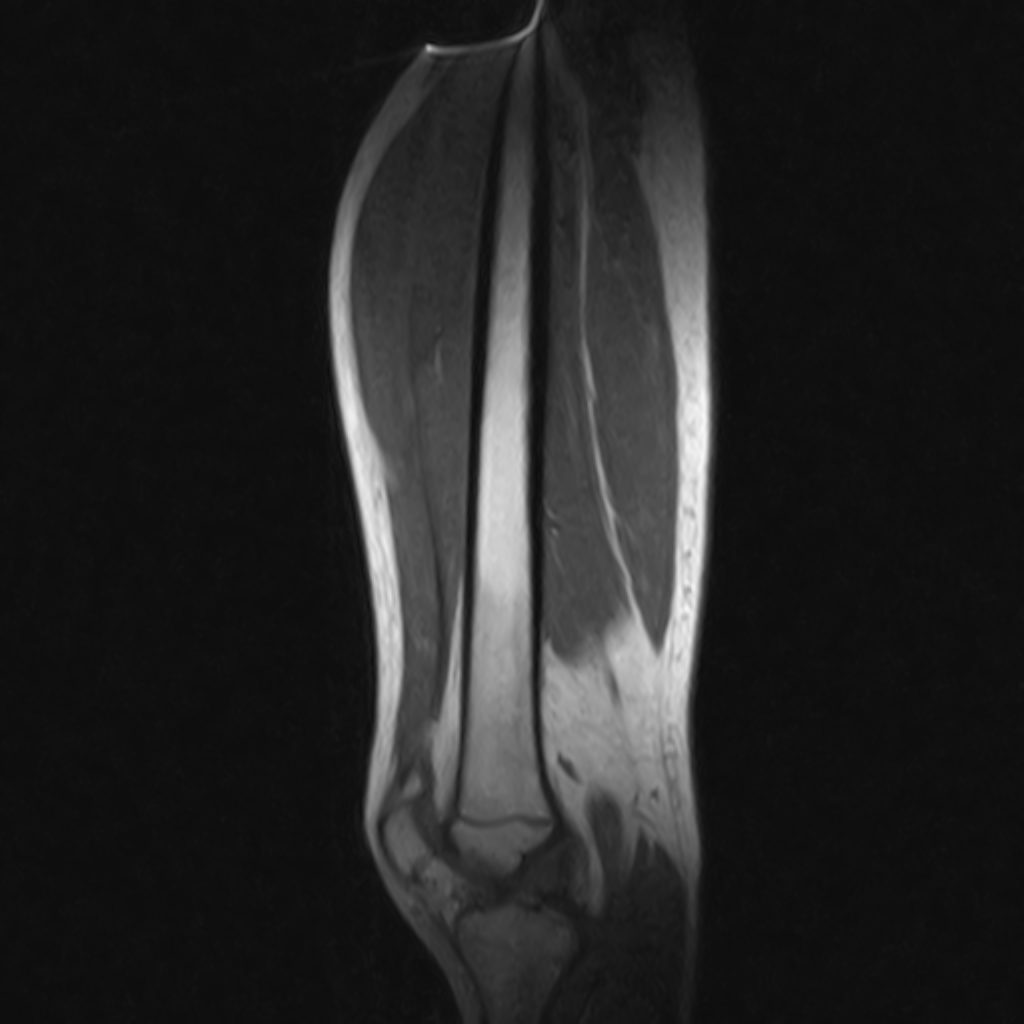
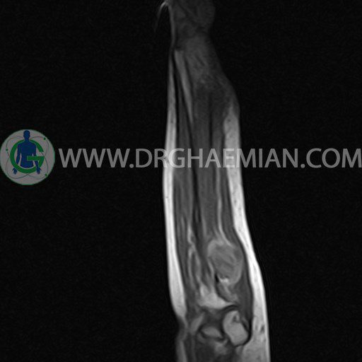
ام آر آی پارگی جزئی ران پا بیمار
ام آر آی ران روشی تصویربرداری از ران پا است که با استفاده از آهنرباهایی قوی تصاویری از پا ایجاد…
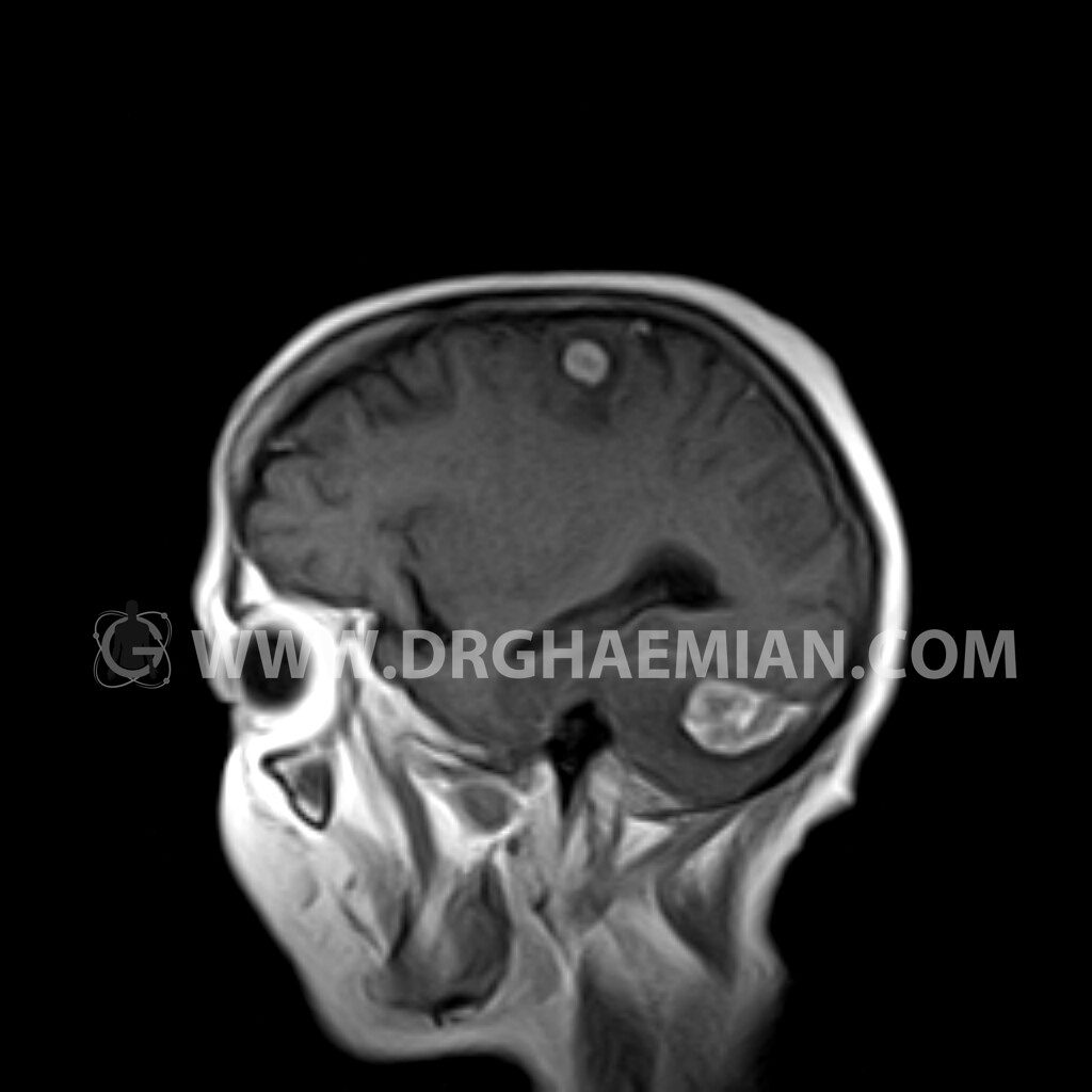
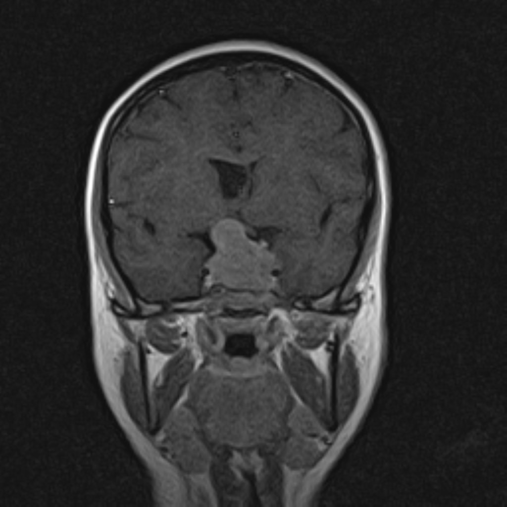
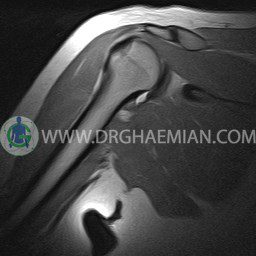
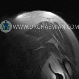
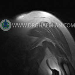
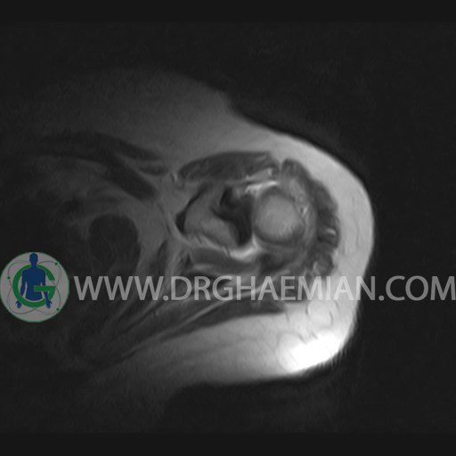
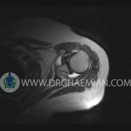
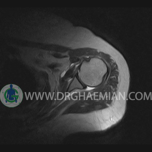
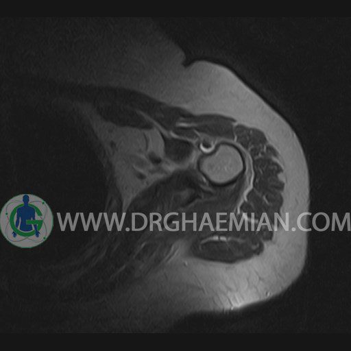
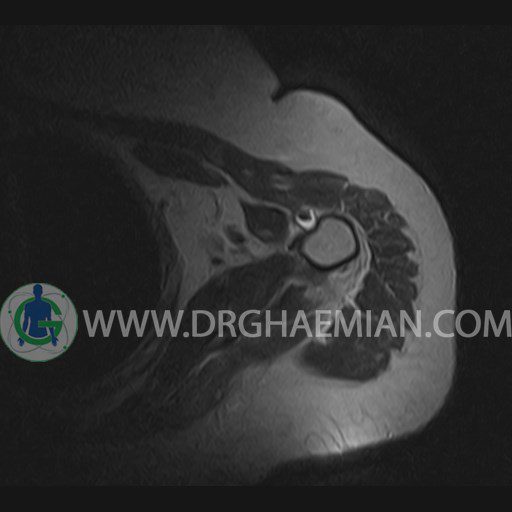
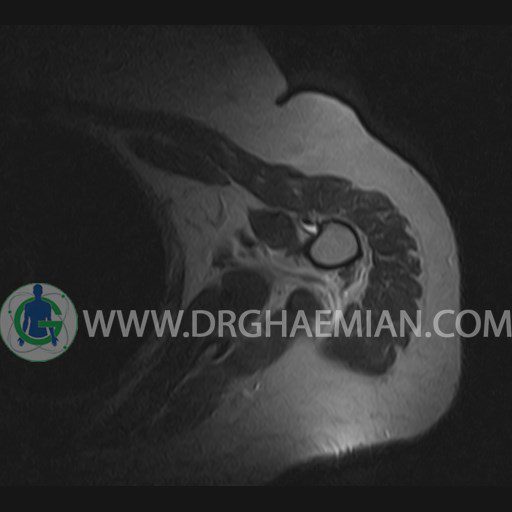
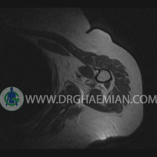
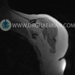
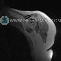
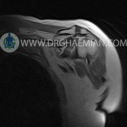
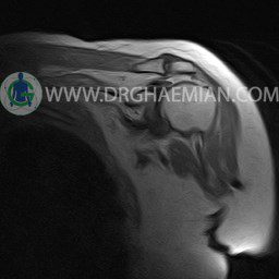
ام آر آی آتروفی عضلانی کتف بیمار
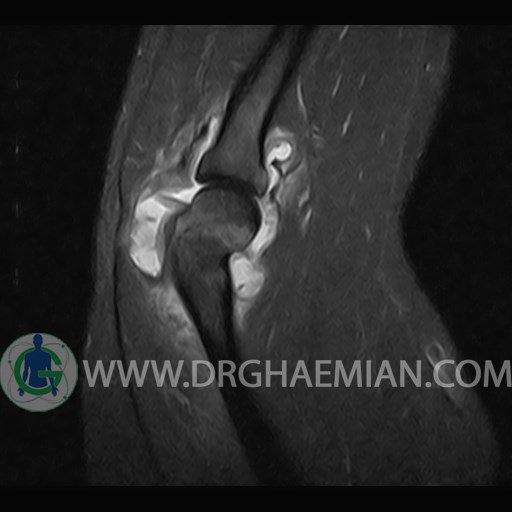
ام ار آی کتف یک روش تصویربرداری است که به وسیله آهنرباهای قدرتمند از قسمت کتف تصاویری ایجاد می کند. این نوع تصویربرداری از تشعشعات استفاده نمی کند. در این کیس آتروفی عضلانی کتف به همراه پارگی تاندون بالاخاری، بورسیت ساب دلتوئید و افیوژن مفصل دیده می شود.
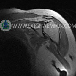


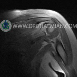


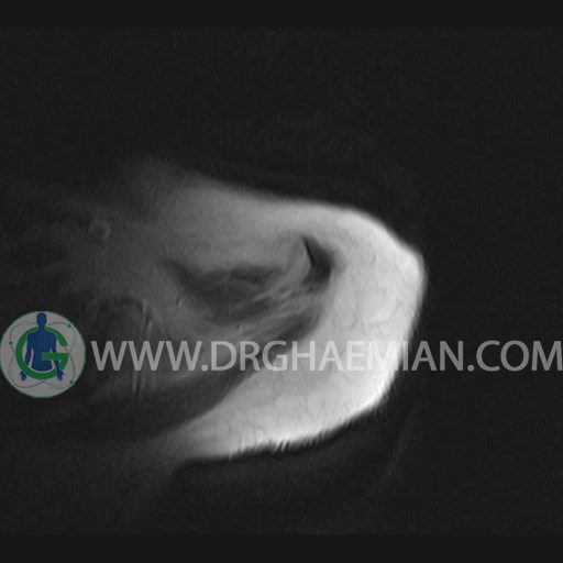
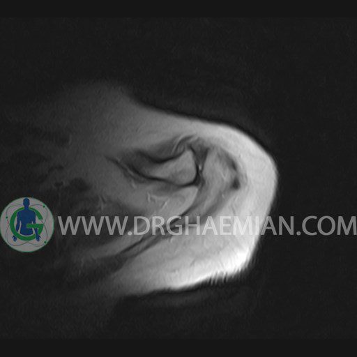


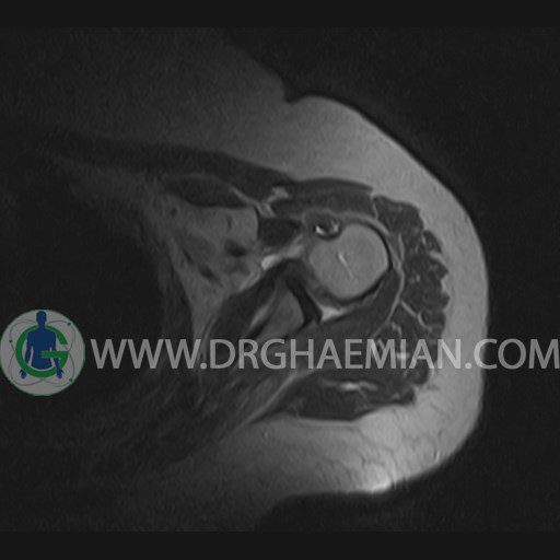


گزارش پزشک :
LEFT SHOULDER MRI
(Without contrast)
Technique: Axial T1 and GE , coronal T1 , T2 , sagittal T1 , T2 .
REPORT:
The humeral head has normal configuration and articulates properly and parallelism with the normally developed glenoid .
The articular surfaces are smooth and show normal cortical thickness .
The bone marrow displays homogeneous , fat – equivalent signal intensity .
The glenoid labrum is intact on all sides .
The biceps tendon appears normal and occupies a normal position in the bicipital groove.
The other muscles that cover the shoulder joint appear normal , as do imaged portions of the lungs and soft tissues .
– Complete tearing of supraspinatus tendon with grade 2 retraction and muscle atrophy
– AC joint hypertrophy with subacromial – subdeltoid bursitis
– Glenohumeral joint effusion
are seen.