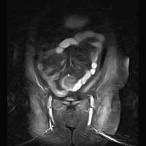
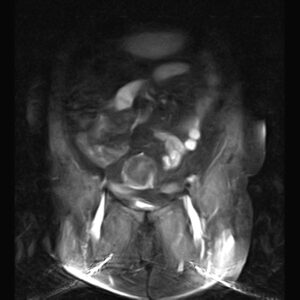
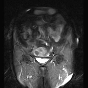
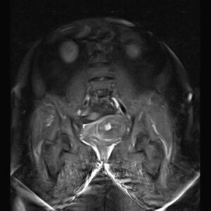
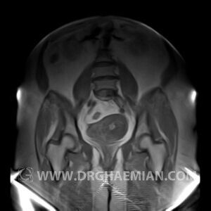
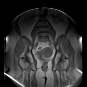
ام آر آی لگن یک روش تصویربرداری است که از طریق دستگاهی با آهنرباهایی قوی و امواج رادیویی از ناحیه بین استخوان های ران تصاویری می سازد. این قسمت از بدن را ناحیه لگنی می گویند. ساختار داخل و پیرامونی لگن شامل مثانه، پروستات، و دیگر اندام های تولید مثل مذکر، اندام های تولید مقل مونث، غدد لنفاوی، روده بزرگ، روده کوچک و استخوان های لگنی می شود. ام آر آی لگن از تششعات استفاده نمی کند.
دلایل تجویز ام آر آی لگن
این نوع تصویربرداری برای بانوان زمانی انجام می شود که نشانه یا علائم زیر را داشته باشند:
- خونریزی غیرطبیعی واژن
- وجود توده در لگن (لمس شده هنگام معاینه لگن یا دیده شده در نوعی دیگر از تصویربرداری)
- فیبروم
- توده ی لگنی که در دوره بارداری ایجاد می شود
- ذخیم شده دیواره رحم (معمولا بعد از سونوگرافی انجام می شود)
- درد در ناحیه پایینی شکم
- ناباروری غیر قابل توضیح (معمولا بعد از سونوگرافی انجام می شود)
- درد بدون دلیل در لگن (معمولا بعد از سونوگرافی انجام می شود)
HIP & RIGHT THIGH MRI
( with & without contrast )
REPORT:
The femoral heads and acetabula are normal shape , signal intensity and the femoral heads are well covered by the acetabular margins .
The joint spaces are of normal width without fluid collection .
the articuler surfaces are smooth and congruent and show normal cortical thickness .
The bone marrow shows normal signal intensity , especially in the femoral head and neck .
– A larga fat signal mass lesion ( 65 x 70 x 150 mm ) in anterior of proximal thigh between rectus femoris & intermedius of quadriceps muscle , without post contrast enhancement suggestive for lipoma
– Intra – mural myoma (14 mm ) in uterin fondus
PELVIC MRI
( with & without contrast)
REPORT:
The pelvic inlet appears normal , with normal configuration of iliac wings and iliopsoas muscles.
No abnormalities are found in imaged bowel structures and there are no signs of wall thickening or mass lesions.
The adnexa appear normal on both sides.
The urinary bladder appears normal and has normal wall thickness.
The femoral heads are normally shaped and articulate normally with the acetabula they have normal bone marrow signal characteristics.
The muscles around the pelvis are unremarkable.
Sacroiliac joints are unremarkable.
– Multiple intra mural – subserosal myoma :
1. ( 40 x 45 mm ) in right side of fondus
2. ( 20 x 22 mm ) & ( 20 x 25 mm ) in right side of body
3. ( 48 x 55 mm ) in left side of body
– Both ovaries are atrophied & without follicles .

