ام آر آی کف پا و انگشتان یک روش تصویربرداری پزشکی است که با استفاده از میدانهای مغناطیسی قوی و امواج رادیویی تصاویر دقیق و باکیفیتی از ساختارهای داخلی کف پا و انگشتان ایجاد میکند. این روش بدون استفاده از اشعههای مضر مانند اشعه ایکس انجام میشود و به پزشک کمک میکند تا با جزئیات بیشتری آسیبها، التهابها یا مشکلات دیگر را تشخیص دهد.در ان کیس یک پای بیمار مبتلا بهب بیماری دیابت دیده میشود.

گزارش پزشک
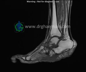
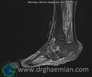
MRI OF RIGHT FOOT
1.5 Tesla MR System
Multislice, multiplanar and multisequence MR images findings:
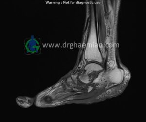
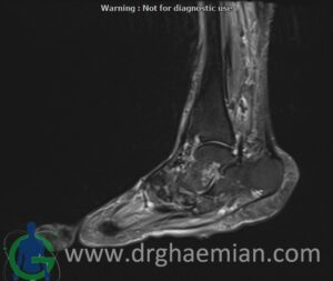
There is a skin ulcer in the plantar aspect of the foot, superficial to the midfoot, without obvious collection.
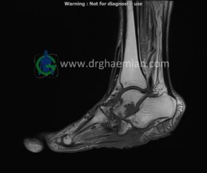
Destruction and erosion of the midfoot structures and articular spaces are seen, resulting in midfoot collapse.
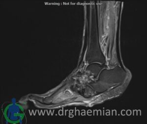
T1 signal intensity of bone marrow in the midfoot region is preserved, and there is no imaging evidence of acute osteomyelitis; however, chronic osteomyelitis cannot be ruled out.
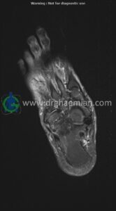
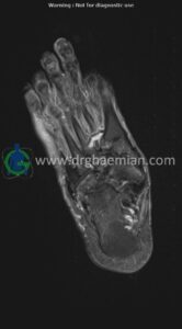
Abnormal high T2WI and low T1WI signal intensity are observed in the 5th proximal and distal phalanges, in association with a suspicious plantar site ulcer, highly suggestive of osteomyelitis in this area.
Other bones, joints, and soft tissues of the foot appear unremarkable.
Impression:
Plantar skin ulcer overlying the midfoot with no associated collection
Structural destruction and erosion of midfoot bones and articular spaces causing midfoot collapse, without definite signs of acute osteomyelitis, though chronic osteomyelitis cannot be excluded
Abnormal marrow signal changes in the 5th proximal and distal phalanges in association with a suspicious plantar ulcer, highly suggestive of osteomyelitis
No other abnormality detected
