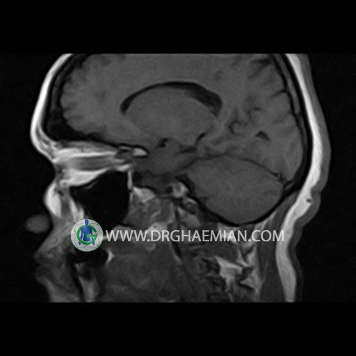
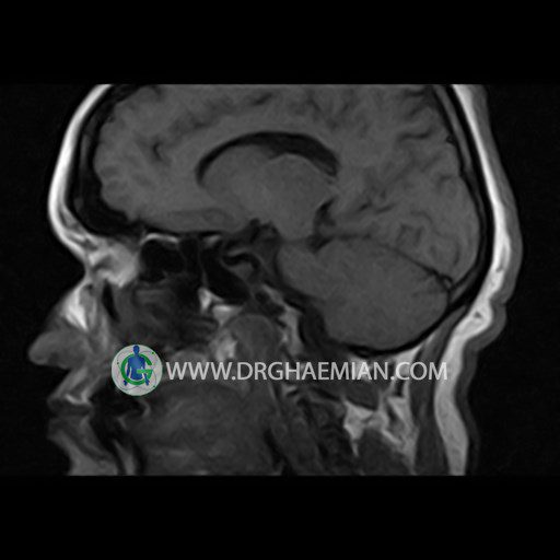
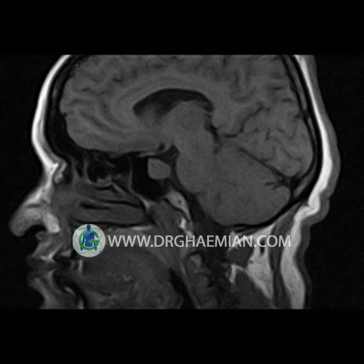
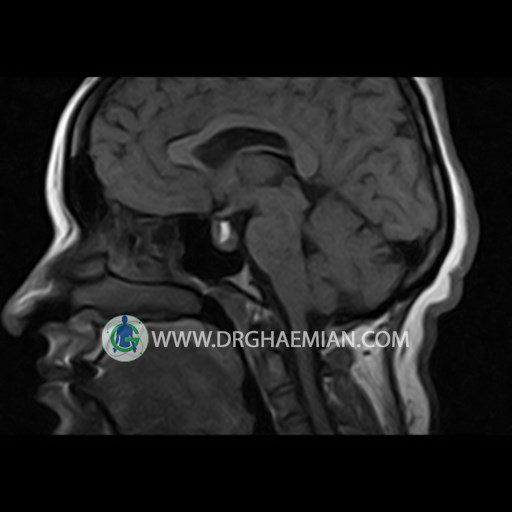
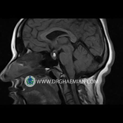
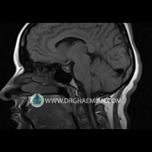
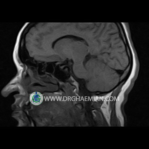
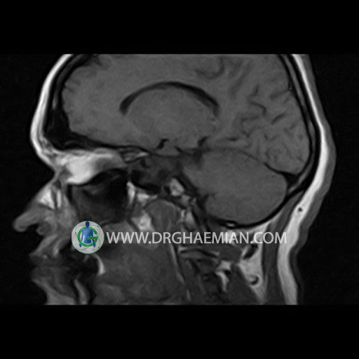
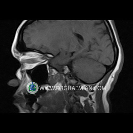
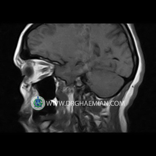
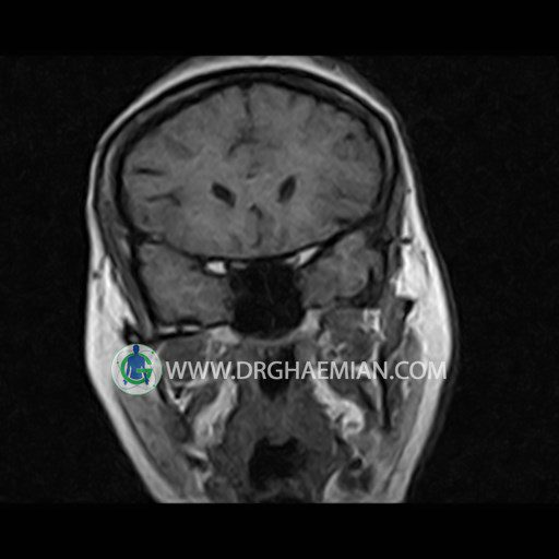
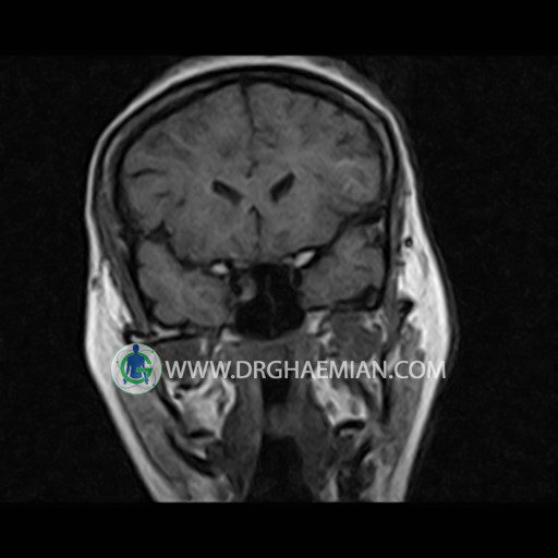
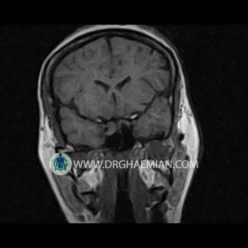
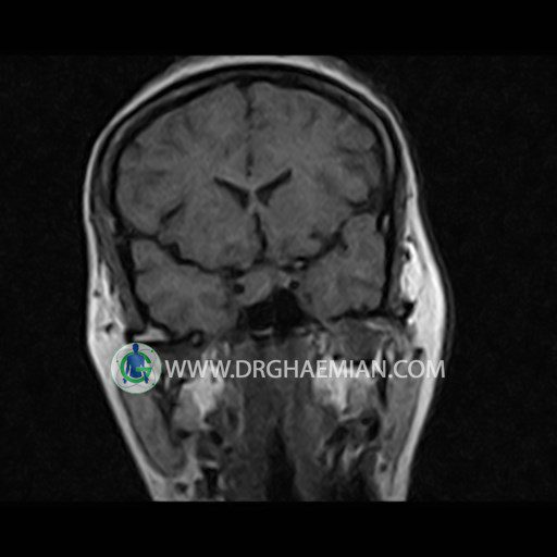
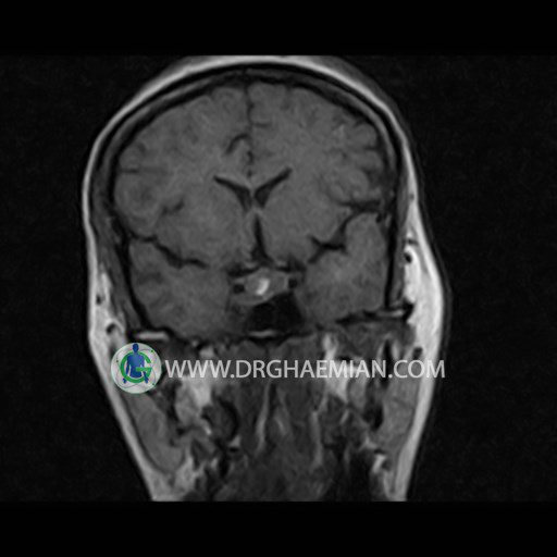
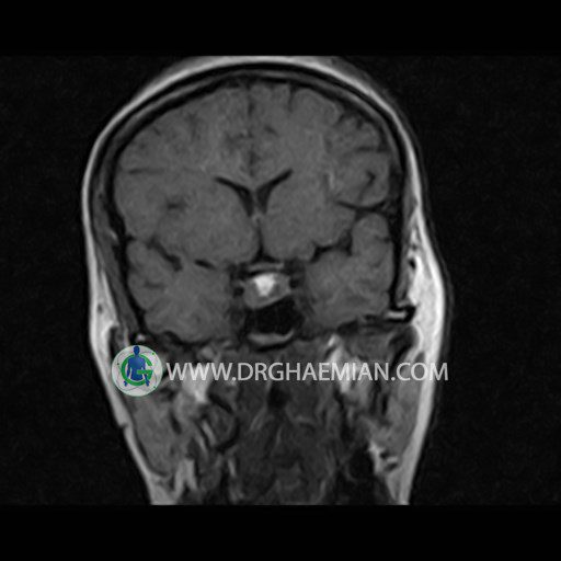
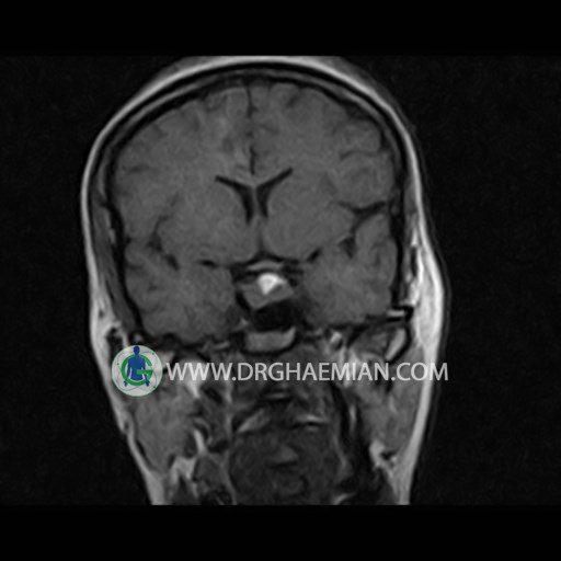
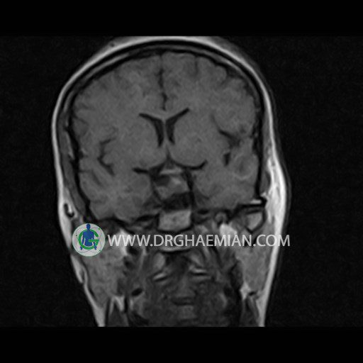
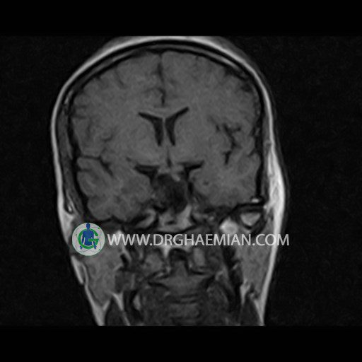
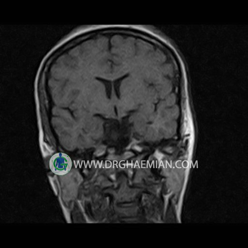
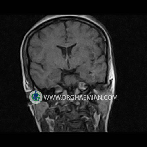
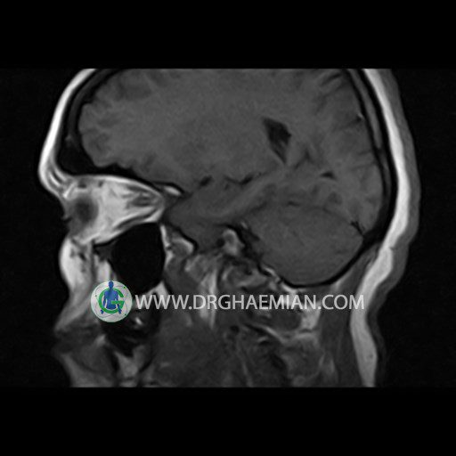
پزشکان اغلب از تصویربرداری ام آر آی برای تشخیص و درمان عارضه های پزشکی که فقط با استفاده از اشعه ایکس یا میدان مغناطیسی و امواج رادیویی قابل مشاهده است، استفاده می کنند. دستگاه ام آر آی تصاویر دقیق از ساختار های داخلی بدن ایجاد می کند. در این کیس میکروآدنوم با دژنراسیون کیستیک و خونریزی مشاهده می شود.
گزارش پزشک :
HYPOPHYSIS MRI
(with and without contrast)
Technique: Axial , coronal T1 , Axial , coronal , sagittal T2 , Axial, coronal T1 post Gd & 64 dynamic thin coronal slices.
REPORT :
The sella shows normal size , position and configuration .
The borders of its floor and walls are smooth and sharply defined .
The infundibulum is centered and of normal size .
The optic chiasm and suprasellar spaces appear normal .
The cavernous sinus and imaged portions of the internal carotid artery and carotid siphon are unremarkable .
Evaluable portions of the neurocranium show no abnormalities .
The sphenoid sinus is clear and pneumatized .
Imaging of the hypothalamus after contrast medium administration was normal.
– A well – defined mass like lesion ( 8 x 9 mm ) in central portion of pituitary gland ( intermediate T1 , high T2) suggestive for pituitary microadenoma with cystic degeneration & hemorrhage
is seen
