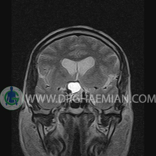
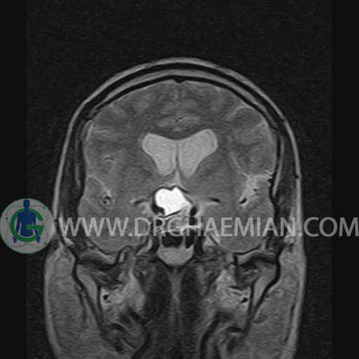
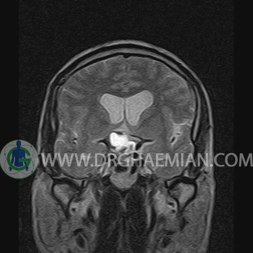
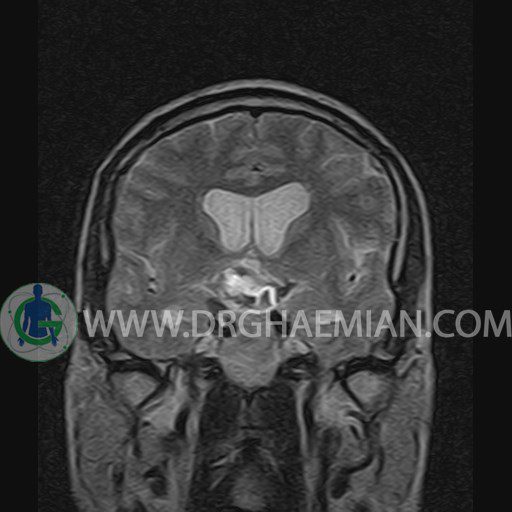
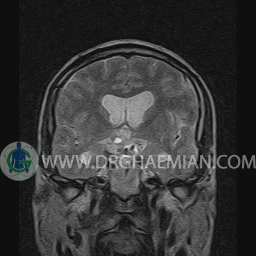
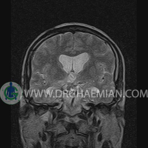
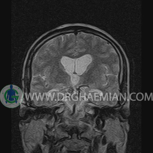
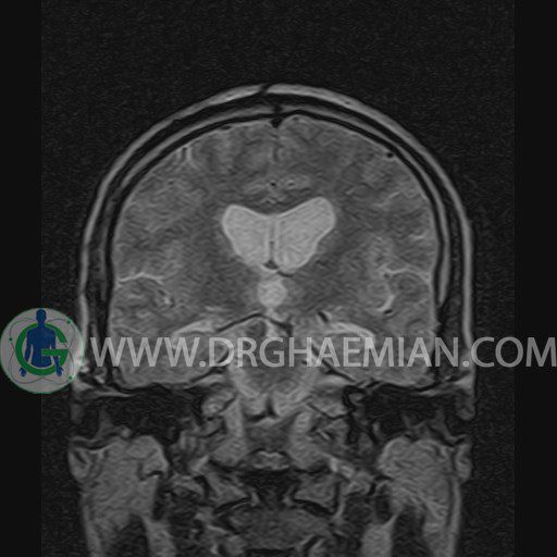
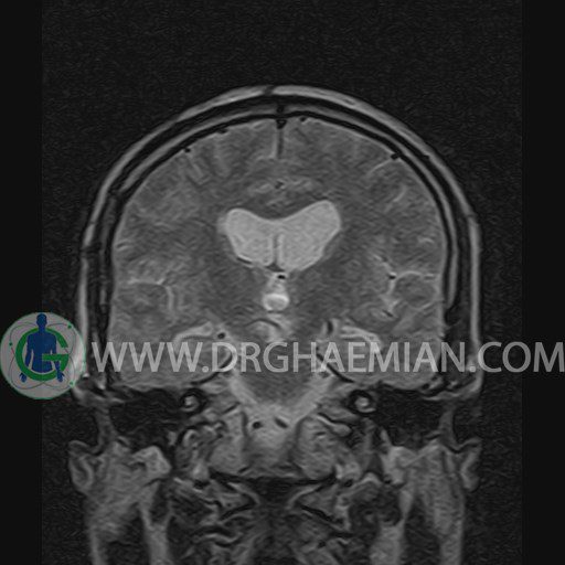
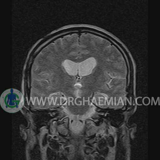
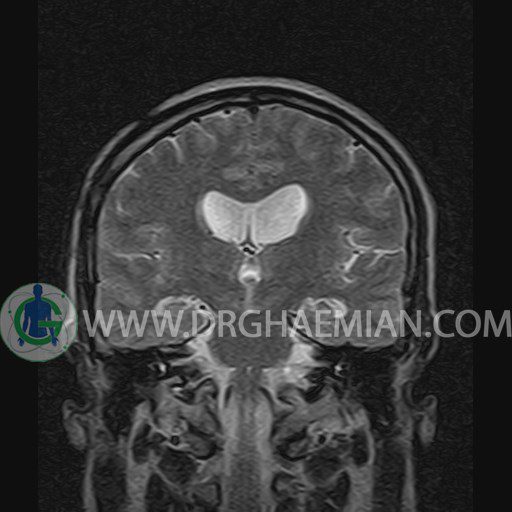
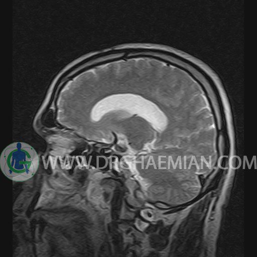
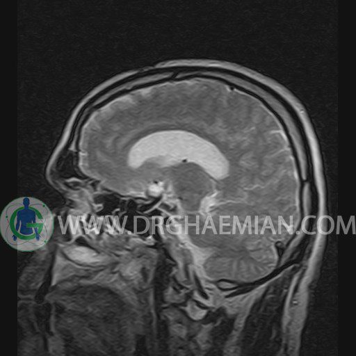
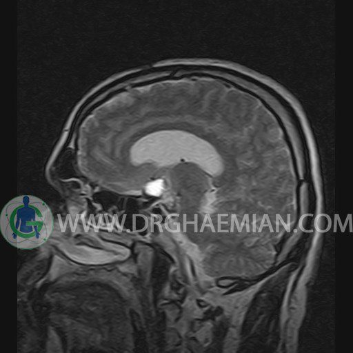
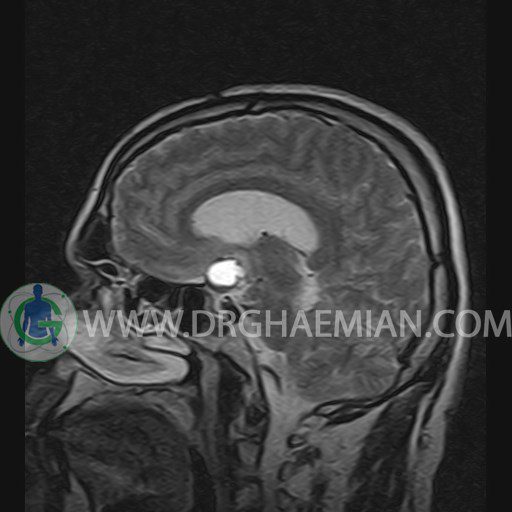
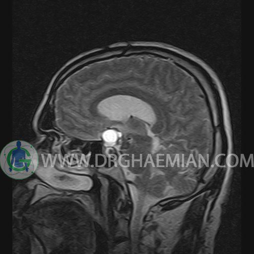
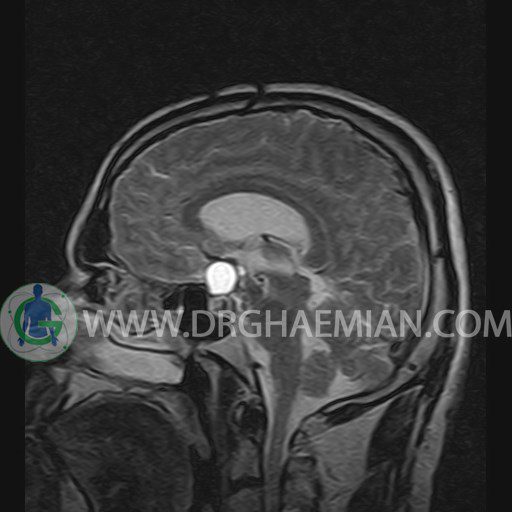
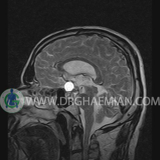
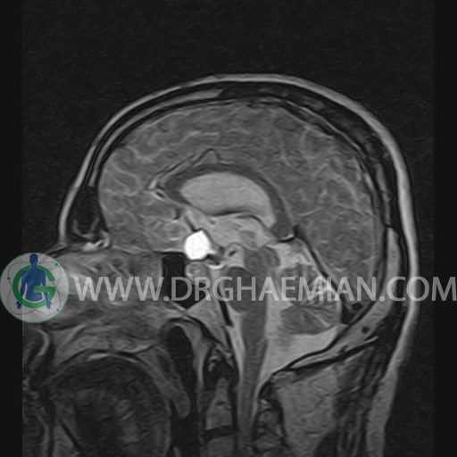
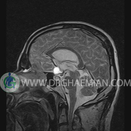
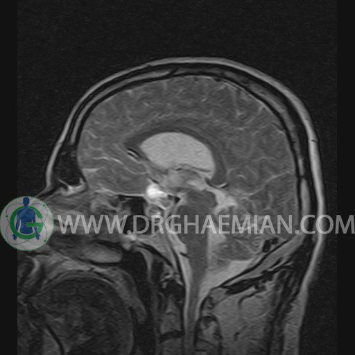
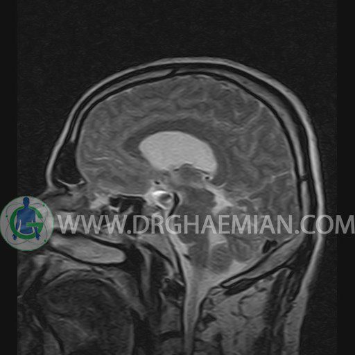
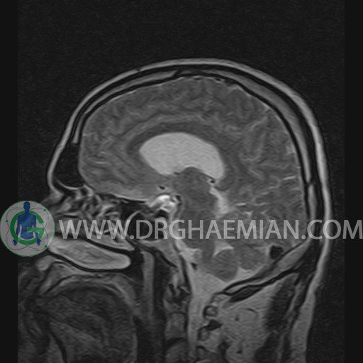
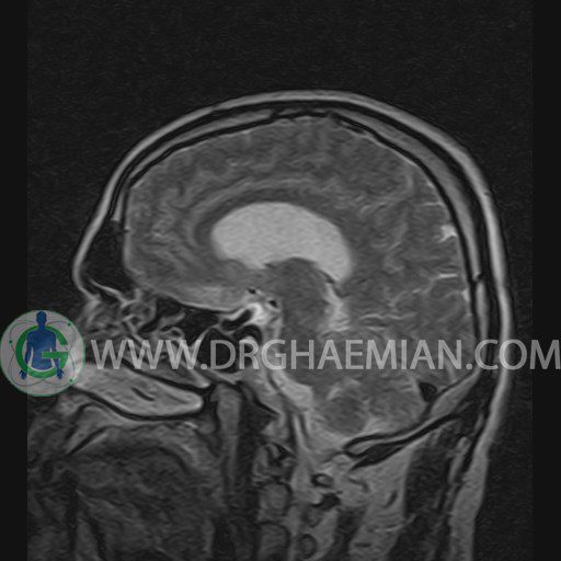
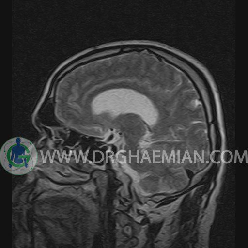
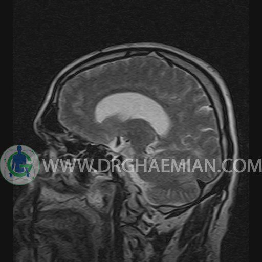
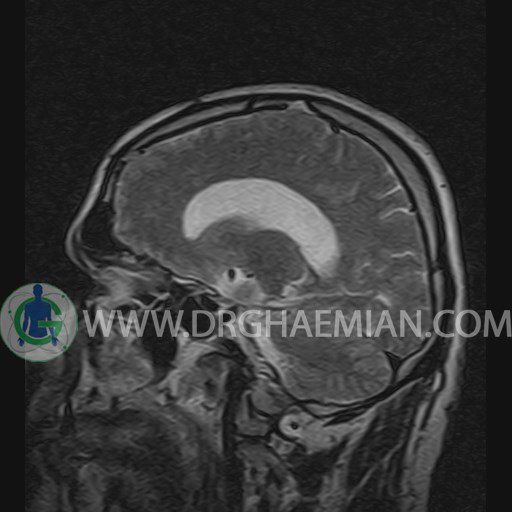
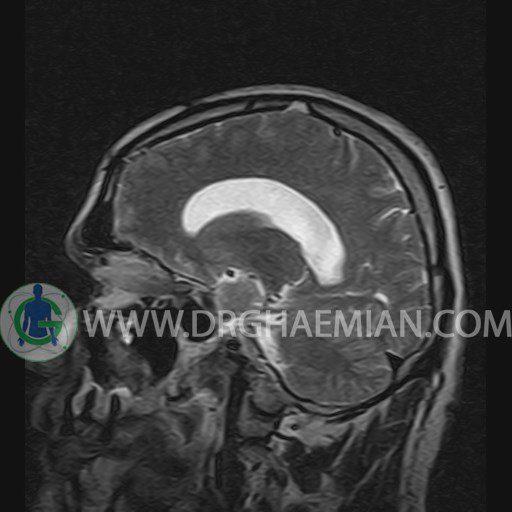
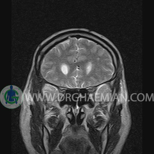
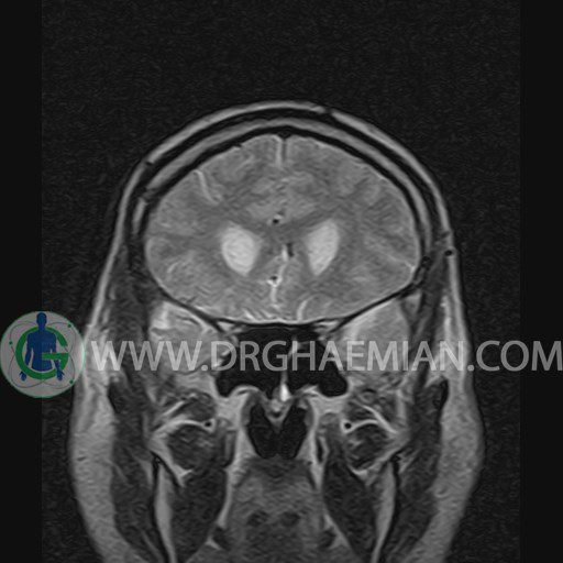
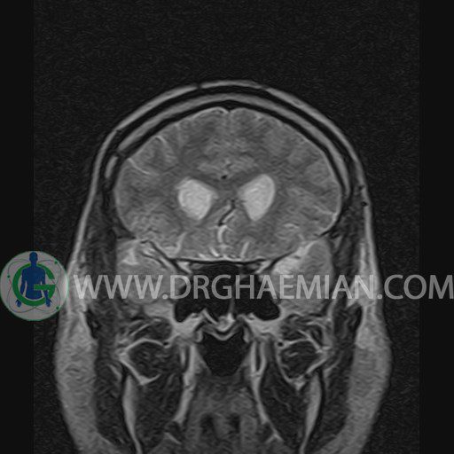
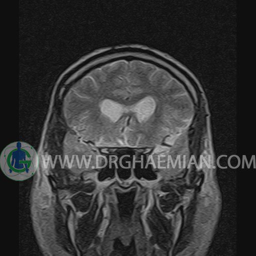
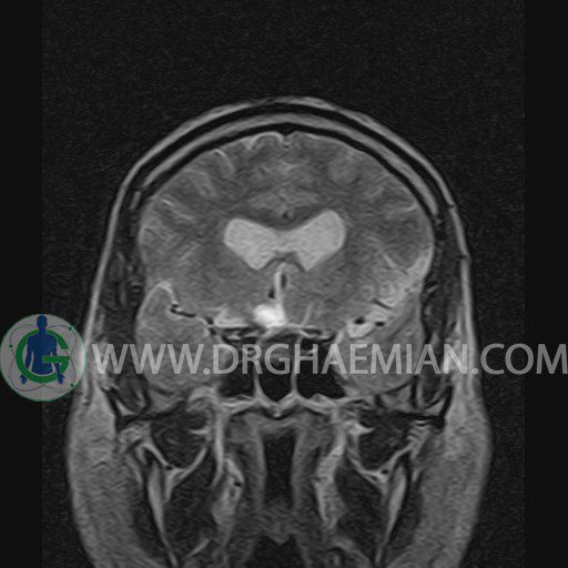
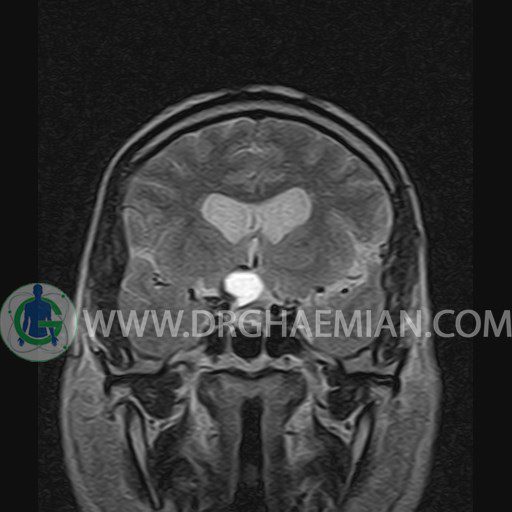
پزشکان اغلب از تصویربرداری ام آر آی برای تشخیص و درمان عارضه های پزشکی که فقط با استفاده از اشعه ایکس یا میدان مغناطیسی و امواج رادیویی قابل مشاهده است، استفاده می کنند. دستگاه ام آر آی تصاویر دقیق از ساختار های داخلی بدن ایجاد می کند. در این کیس کرانیوفارنژیوما به ابعاد mm 15 x 27 و کرانیوتومی در قسمت جلویی سر دیده می شود.
گزارش پزشک :
HYPOPHYSIS MRI
(with and without contrast)
Technique: Axial , coronal T1 , Axial , coronal , sagittal T2 , Axial, coronal T1 post Gd & 64 dynamic thin coronal slices.
REPORT :
The pituitary tissue shows normal , position, shape , size and homogeneous signal intensity both before and after contrast administration .
Anterior and posterior pituitary gland were normal .
The infundibulum is centered and of normal size .
The optic chiasm and suprasellar spaces appear normal .
The cavernous sinus and imaged portions of the internal carotid artery and carotid siphon are unremarkable .
Evaluable portions of the neurocranium show no abnormalities .
The sphenoid sinus is clear and pneumatized .
– Right frontotemporal craniotomy
– A well – defined solid cystic mass lesion ( 15 x 27 mm ) in sella thoracic & suprasellar cistern with post contrast ring enhancement suggestive for recurrent/remnant craniopharyngioma
are seen
