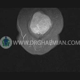
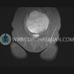
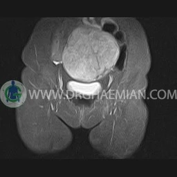
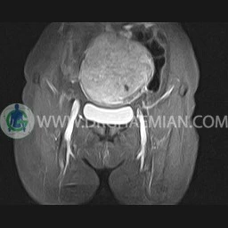
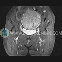
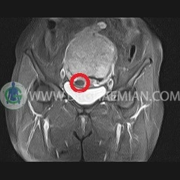
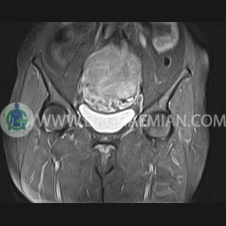
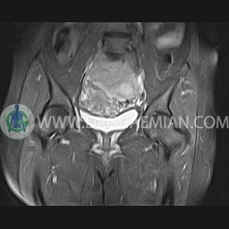
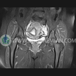
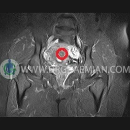
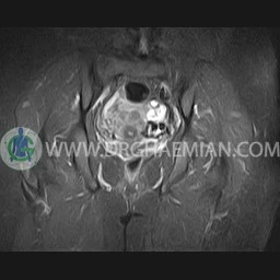
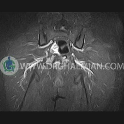
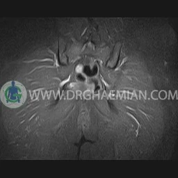
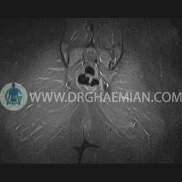
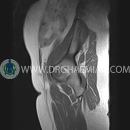
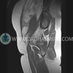
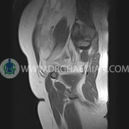
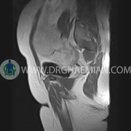
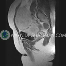
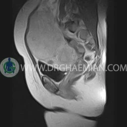
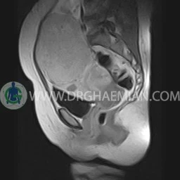
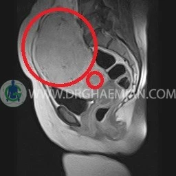
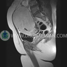
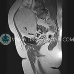
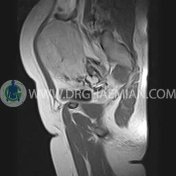
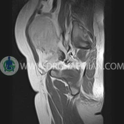
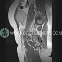
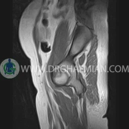
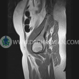
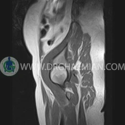
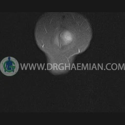
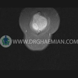
ام ار آی لگن طریق دستگاهی با آهنرباهایی قوی و امواج رادیویی از ناحیه بین استخوان های ران تصاویری می سازد. در این کیس سه میوم به ابعاد mm 95 x 125، mm 12 x 17 و mm 15 x 20 دیده می شود.

گزارش پزشک
PELVIC MRI
( with & without contrast)
Technique : Axial and coronal T2 , coronal T1 & coronal fatsat
REPORT:
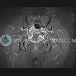
The pelvic inlet appears normal , with normal configuration of iliac wings and iliopsoas muscles.
No abnormalities are found in imaged bowel structures and there are no signs of wall thickening or mass lesions.
The urinary bladder appears normal and has normal wall thickness.
The femoral heads are normally shaped and articulate normally with the acetabula they have normal bone marrow signal characteristics.
The muscles around the pelvis are unremarkable.
Sacroiliac joints are unremarkable.
– Uterin enlargement with :
1. large subserosal pedunculated myoma ( 95 x 125 mm ) attached to fondus
2. intra mural myoma ( 12 x 17 mm ) in anterior of fondus
3. intramural myoma ( 15 x 20 mm ) in left posterior of uterin body
are seen
