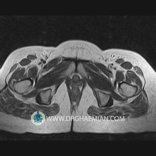
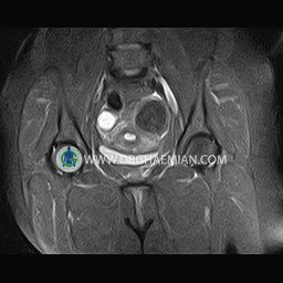
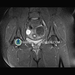
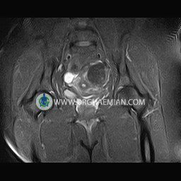
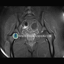
پزشکان اغلب از تصویربرداری ام آر آی برای تشخیص و درمان عارضه های پزشکی که فقط با استفاده از اشعه ایکس یا میدان مغناطیسی و امواج رادیویی قابل مشاهده است، استفاده می کنند. دستگاه ام آر آی تصاویر دقیق از ساختار های داخلی بدن ایجاد می کند. در این کیس میوم ساب سروزال، کیست استخوانی، توده در تخمدان چپ، و کیست در تخمدان راست مشاهده می شود.
گزارش پزشک :
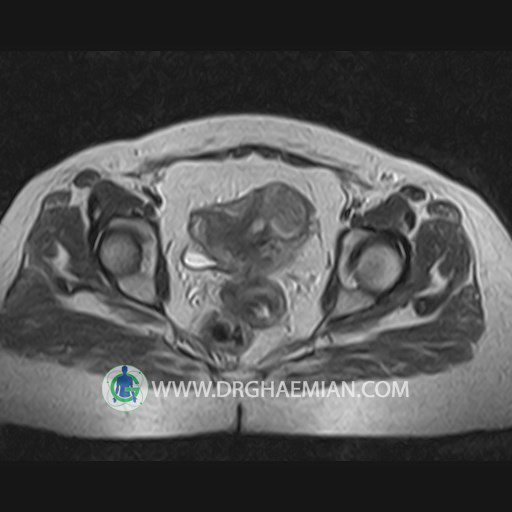
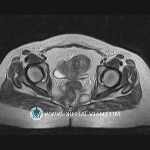
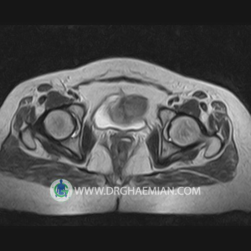
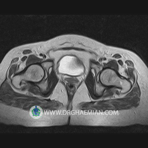
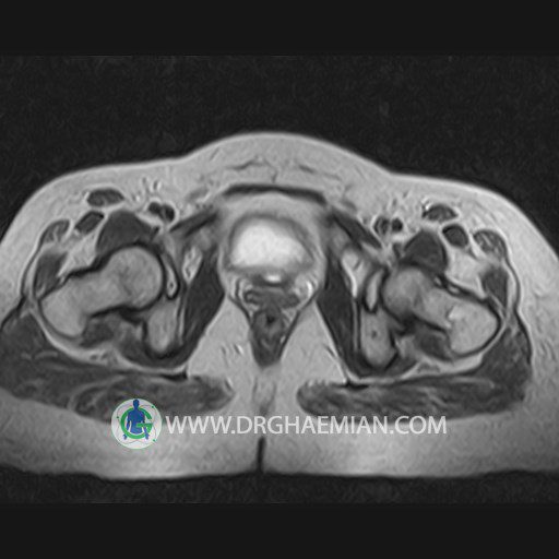
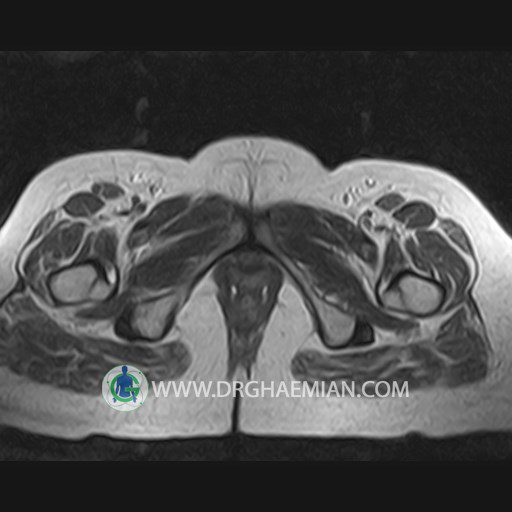
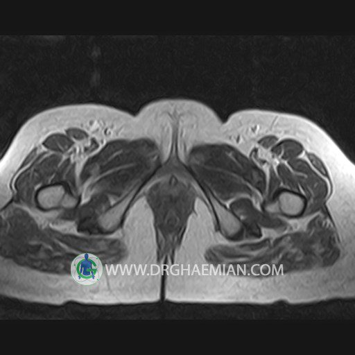
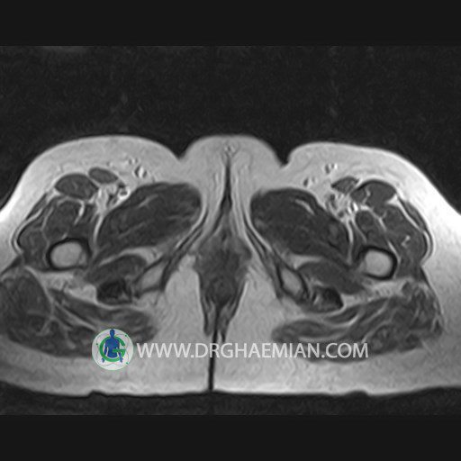
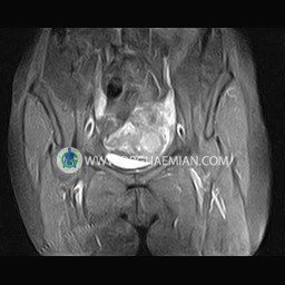
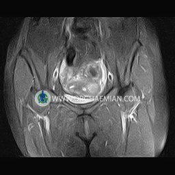
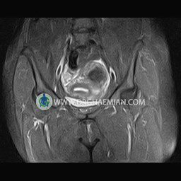
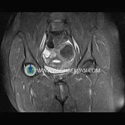
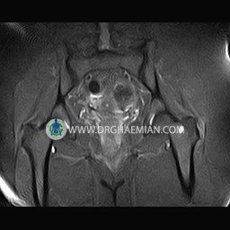
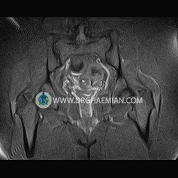
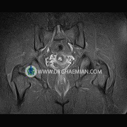
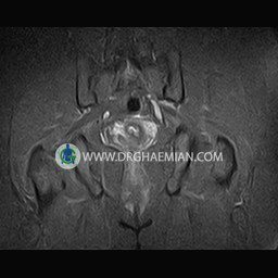
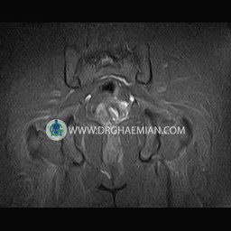
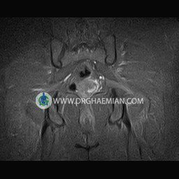
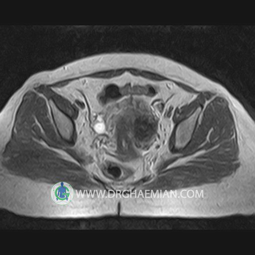
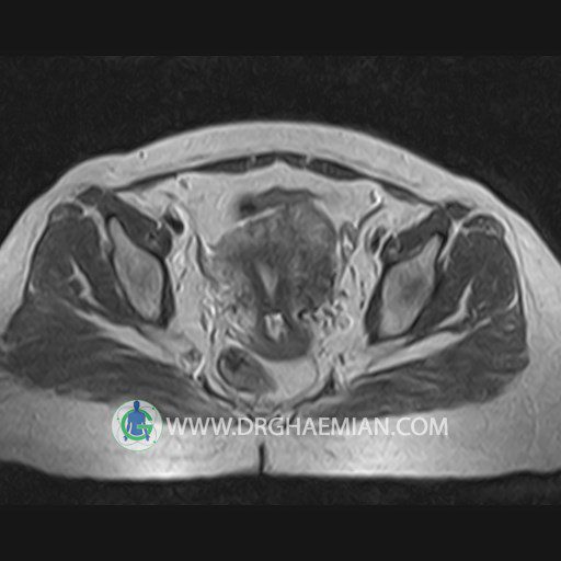
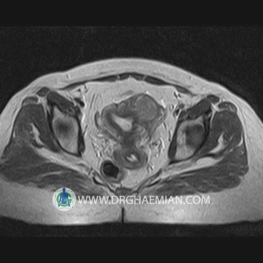
HIP JOINT MRI
( without contrast )
Technique :coronal STIR , coronal T2 , Axial T1 , axial T2 .
REPORT:
The femoral heads and acetabula are normal shape , signal intensity and the femoral heads are well covered by the acetabular margins .
The joint spaces are of normal width without fluid collection .
the articuler surfaces are smooth and congruent and show normal cortical thickness .
The bone marrow shows normal signal intensity , especially in the femoral head and neck .
Each femoral shaft has normal margins and contains a normal bone marrow signal .
The imaged muscles and the lesser pelvis show no abnormalities .
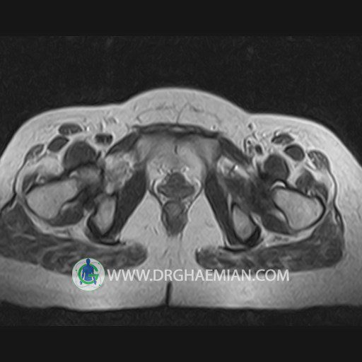
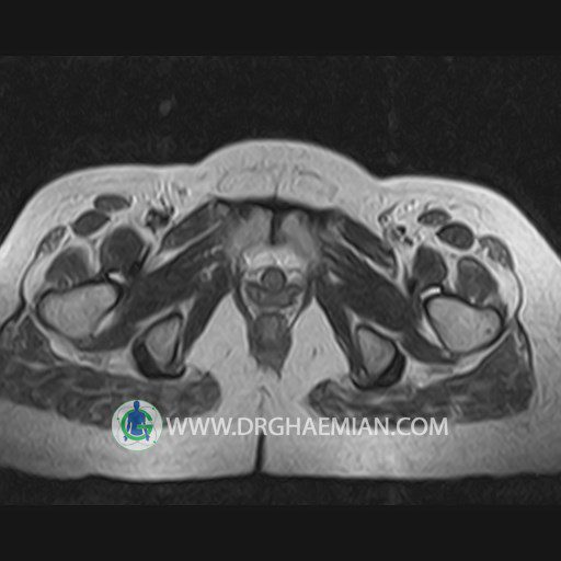
– Focal signal change in trochanteric region of left femur suggestive for bone cyst
– Subserosal myoma ( 28 mm in diameter ) in left side of fondus
– A well – defined mass like lesion in left adnexa ( 45 mm in diameter ) suggestive for left ovarian mass lesion
are seen
INCIDENTAL FINDING : small cyst like lesion in right ovary ( 25 mm )
COMMENT : vaginal sonography is recommended .
