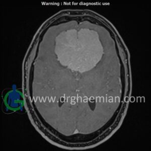
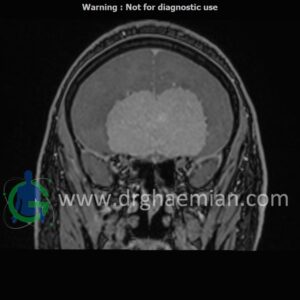
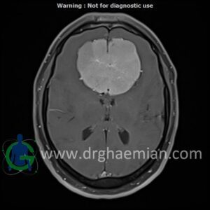
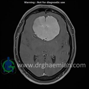
ام آر آی مغز یک روش تصویربرداری است که با استفاده از آهنربا های قوی و امواج رادیویی تصاویری از مغز و بافت های عصبی پیرامونی آن ایجاد می کند. در این کیس مننژیوما مغزی و …. دیده میشود
گزارش پزشک
MRI OF BRAIN (WITH AND WITHOUT IV GADOLINIUM)
1.5 Tesla MR System
Multiplanar, multislice, and multisequence MR images of the brain were obtained before and after IV gadolinium administration. Findings:
Impression
1. Large, vividly enhancing extra-axial mass in the anteriorcranial fossa (49 × 63 × 73 mm) with “spoke-wheel” vessels, CSF cleft, and broad dural contact—findings suggestive of meningioma. 2. Imaging features favor a left olfactory-groove origin, with the lesion wrapping around the anteriorfalx. 3. Extensive frontal lobe vasogenic edema with mass effect effacing the frontal lobes, frontal horns, and the genu of the corpus
callosum, without hydrocephalus. 4. Posteroinferiorextension into the pituitary fossa while remaining separate from the pituitary gland. 5. Close approximation to both supraclinoid internal carotid arteries and contact with the anteriorcerebral arteries along the
posterosuperiorsurface. 6. Findings suspicious forinvasion of the roof of the left ethmoid sinuses (loss of cortical definition). 7. Otherwise unremarkable MRI brain.
