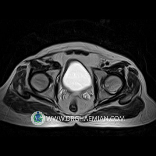
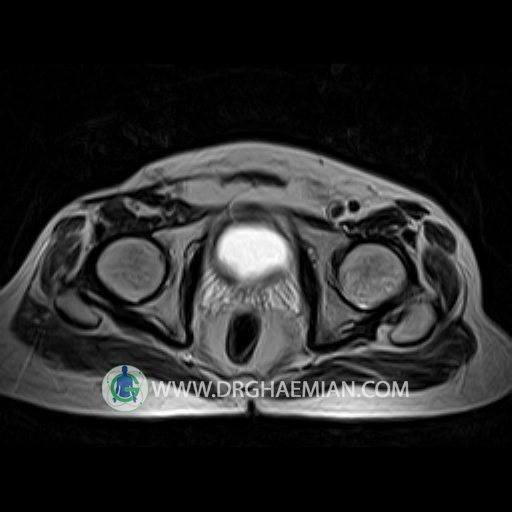
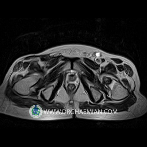
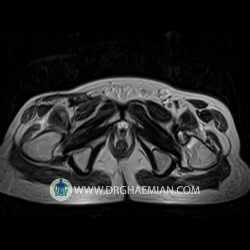
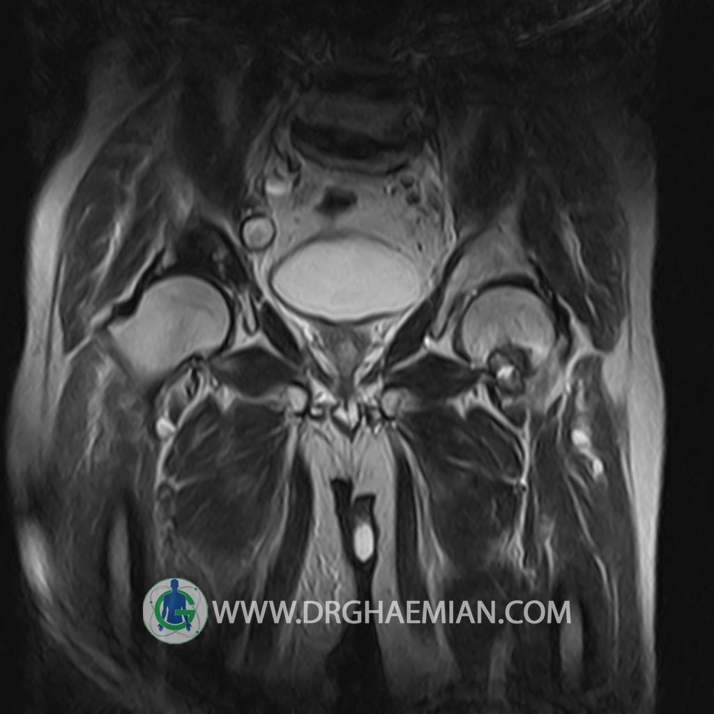
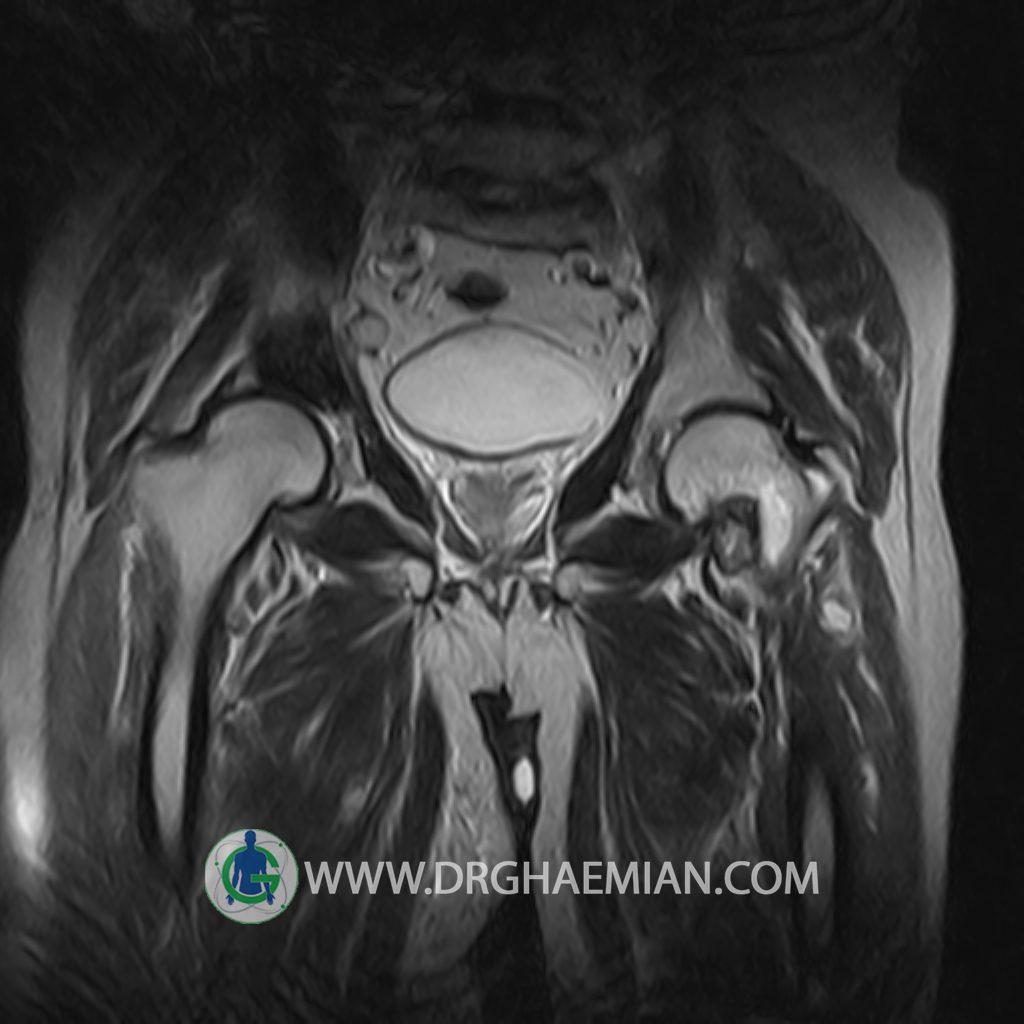
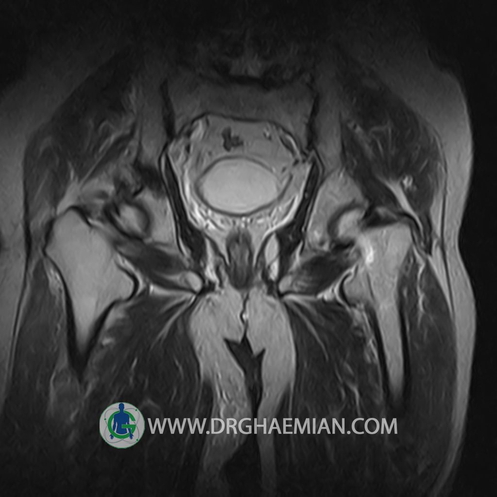
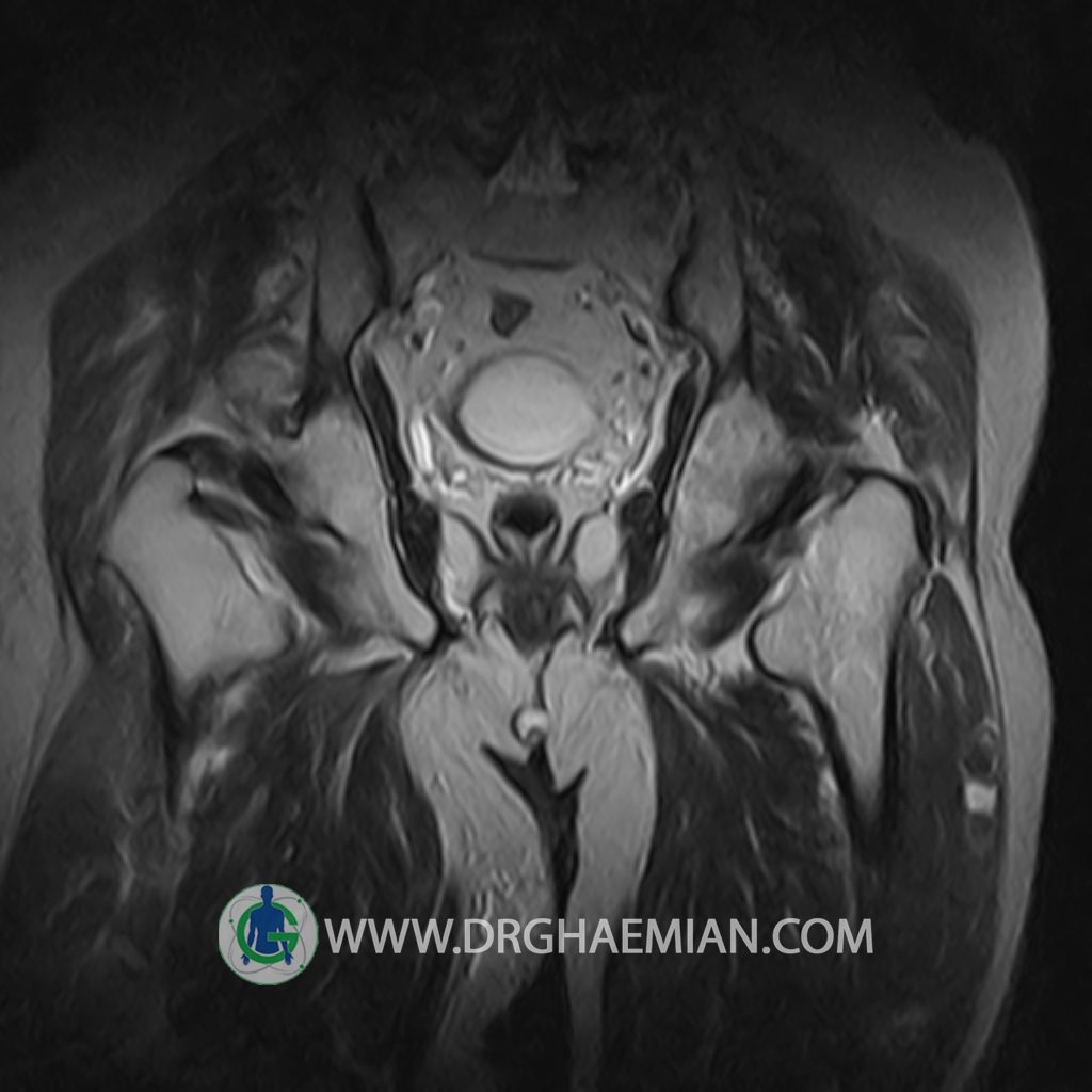
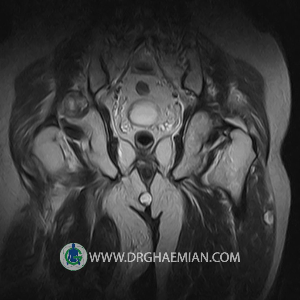
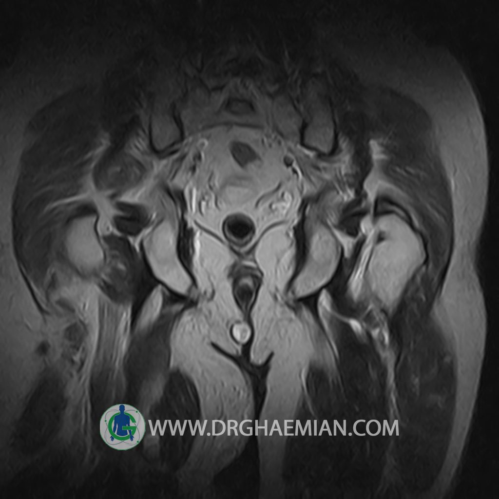
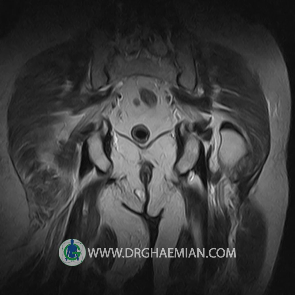
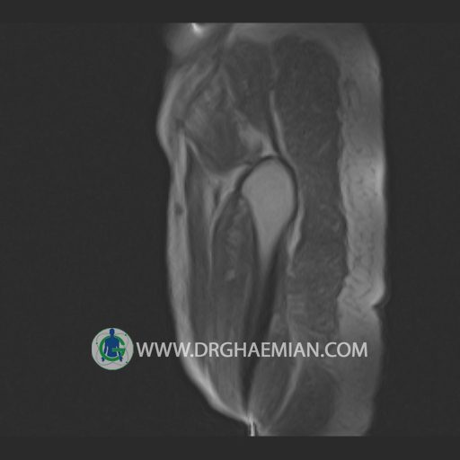
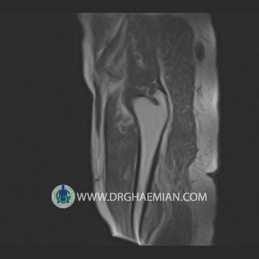
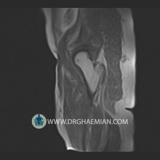
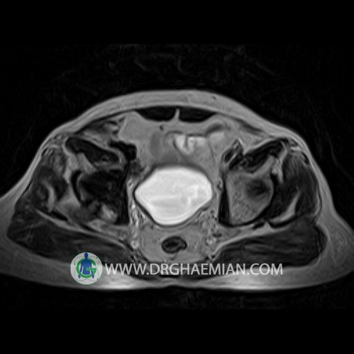
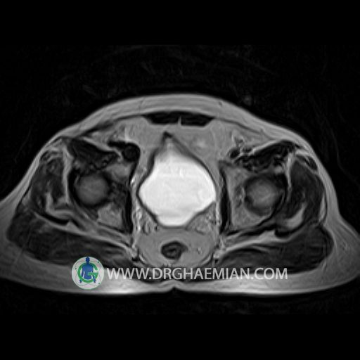
پزشکان اغلب از تصویربرداری ام آر آی برای تشخیص و درمان عارضه های پزشکی که فقط با استفاده از اشعه ایکس یا میدان مغناطیسی و امواج رادیویی قابل مشاهده است، استفاده می کنند. دستگاه ام آر آی تصاویر دقیق از ساختار های داخلی بدن ایجاد می کند. در این کیس تومور استخوانی و متاستاز استخوان لگن تشخیص داده شد.
گزارش پزشک :
PELVIC MRI
(with & without contrast)
Technique : Axial & coronal T1 – axial T2 – coronal STIR & post contrast axial , sagittal , coronal T1 .
REPORT:
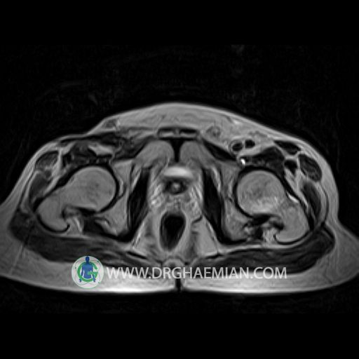
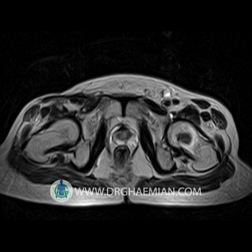
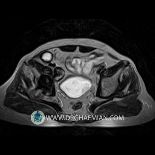
The pelvic inlet appears normal , with normal configuration of iliac wings and iliopsoas muscles.
No abnormalities are found in imaged bowel structures and there are no signs of wall thickening or mass lesions.
The distended urinary bladder normal and has normal wall thickness .
The seminal vesicle are of normal size .
The angle between the bladder and seminal vesicle is normal on each side .
The prostate shows a normal size , configuration and signal intensity .
The vessels of the lesser pelvic are normal in their course and caliber .
There is no evidence of lymphadenopathy .
Sacroiliac joints are unremarcable.
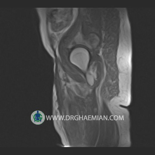
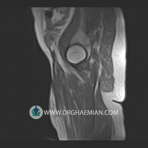
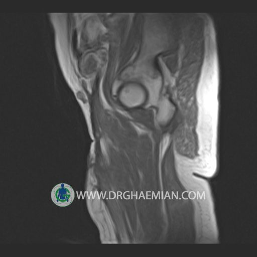
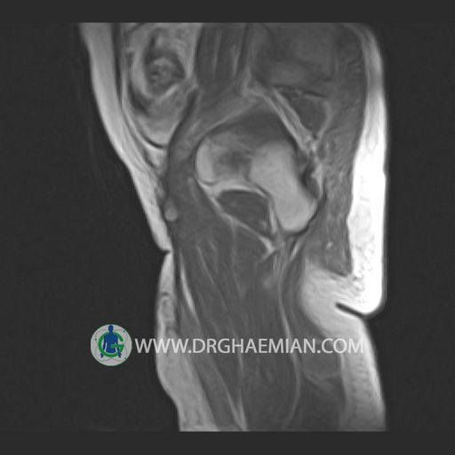
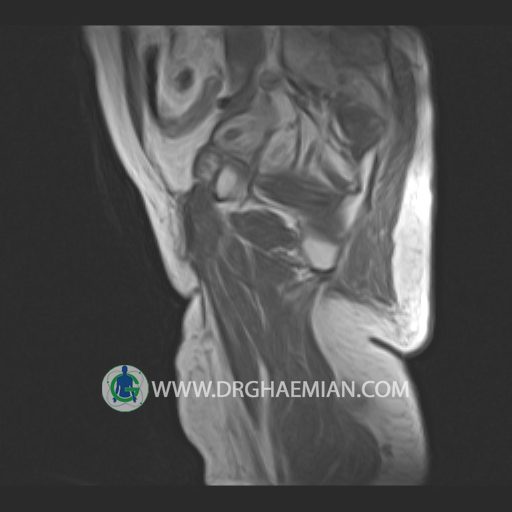
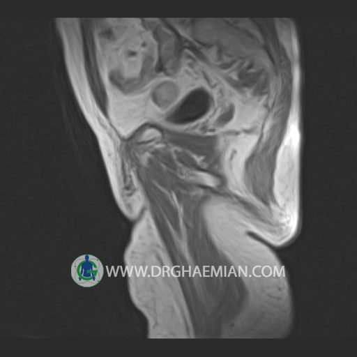
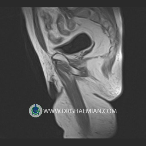
– Heterogeneous signal change in right accetabulum & left femoral neck with soft tissue component & post contrast enhancement suggestive for tumoral infiltration & bone metastasis
is seen
