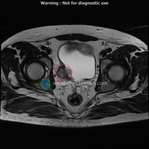
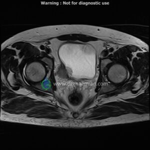
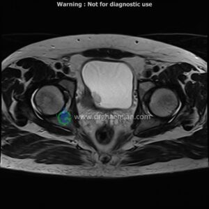
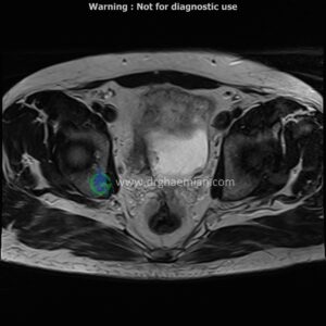
ام آر آی لگن یک روش تصویربرداری است که از طریق دستگاهی با آهنرباهای قوی و امواج رادیویی از ناحیه بین استخوان های ران تصاویری می سازد. این قسمت از بدن را ناحیه لگنی می گویند.در این کیس تومور در ناحیه پایینی دیواره مثانه و … دیده میشود

گزارش پزشک
PELVIC MRI (with & without contrast)
Technique : precontrast :axial T2 haste , axial vibe – dixon – coronal vibe – dixon – sagittal T2/haste – sagittal blade – coronal T2/FATSAT- axial T2-FATSAT- axial DWI with ADC.post contrast :coronal vibe dixon – sagittal vibe dixon – axial vibe dixon – coronal & sagittal T1/FATSAT .
REPORT:
The pelvic inlet appears normal , with normal configuration of iliac wings and iliopsoas muscles.
No abnormalities are found in imaged bowel structures and there are no signs of wall thickening or mass lesions.
The seminal vesicle are of normal size .
The angle between the bladder and seminal vesicle is normal on each side .
The prostate shows a normal size , configuration and signal intensity .
The vessels of the lesser pelvic are normal in their course and caliber .
a patient with urinary bladder mass & surgery .
a wel – defined lobulated heterogeneous mass lesion ( 25x35x38mm ) in right posterior of bladder wall , with extention to perivesical fat , with post contrast peripheral enhancement suggestive for recurrent/remnant tumor
is seen
pelvic lymphadenopathy is not seen .