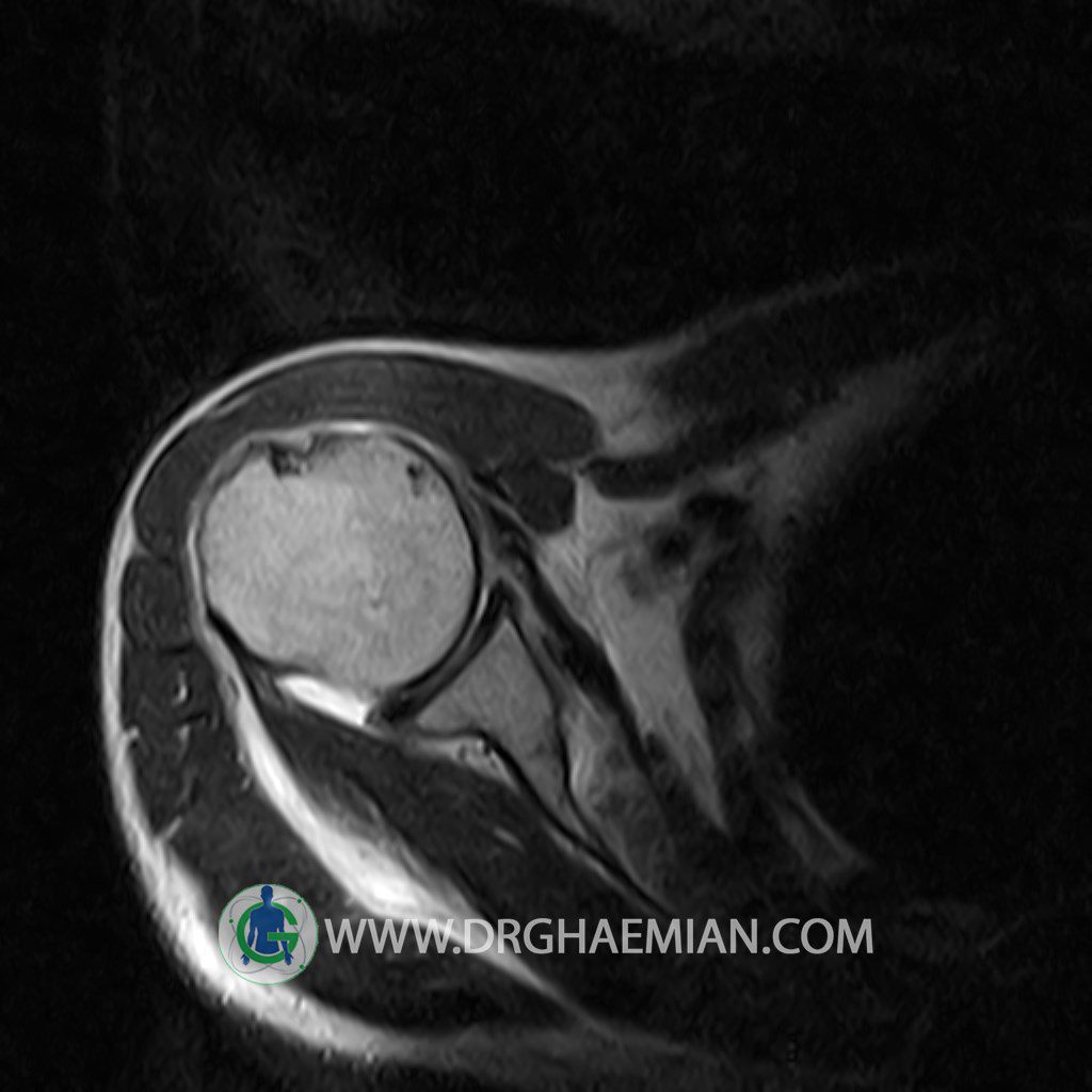
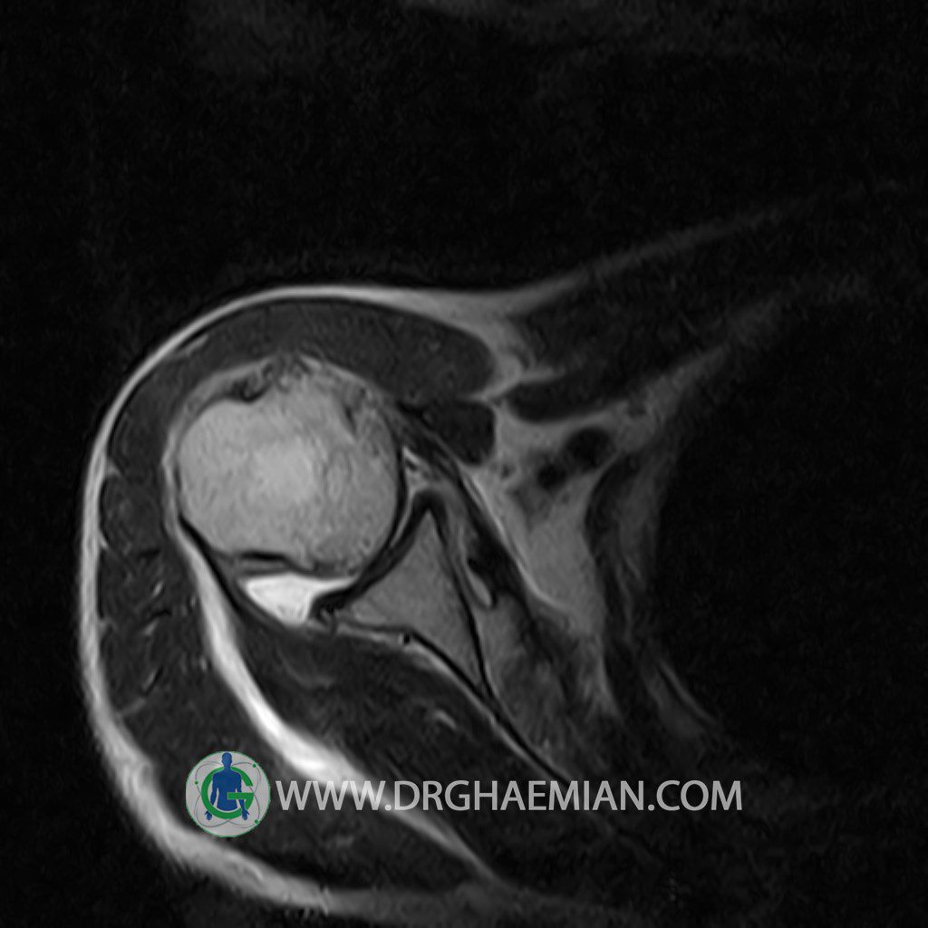
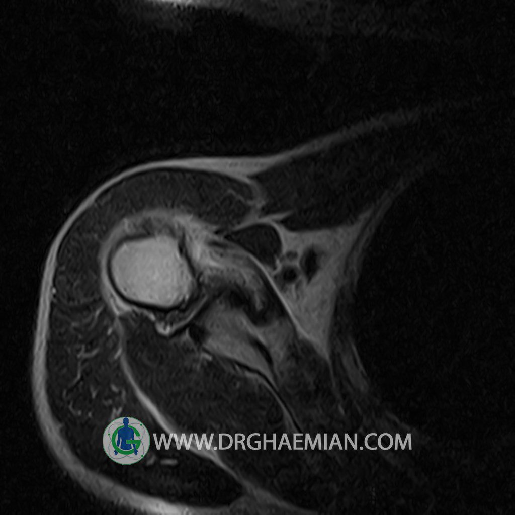
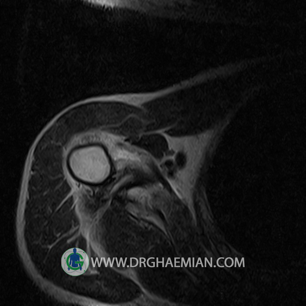
پزشکان اغلب از تصویربرداری ام آر آی برای تشخیص و درمان عارضه های پزشکی که فقط با استفاده از اشعه ایکس یا میدان مغناطیسی و امواج رادیویی قابل مشاهده است، استفاده می کنند. دستگاه ام آر آی تصاویر دقیق از ساختار های داخلی بدن ایجاد می کند. در این کیس بورسیت ساب دلتوئید شانه، پارگی و پیچ خوردگی تاندون و افیوژن مفصل شانه دیده می شود.

گزارش پزشک :
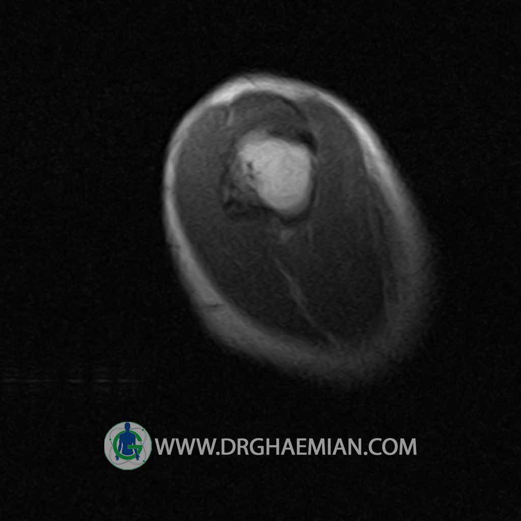
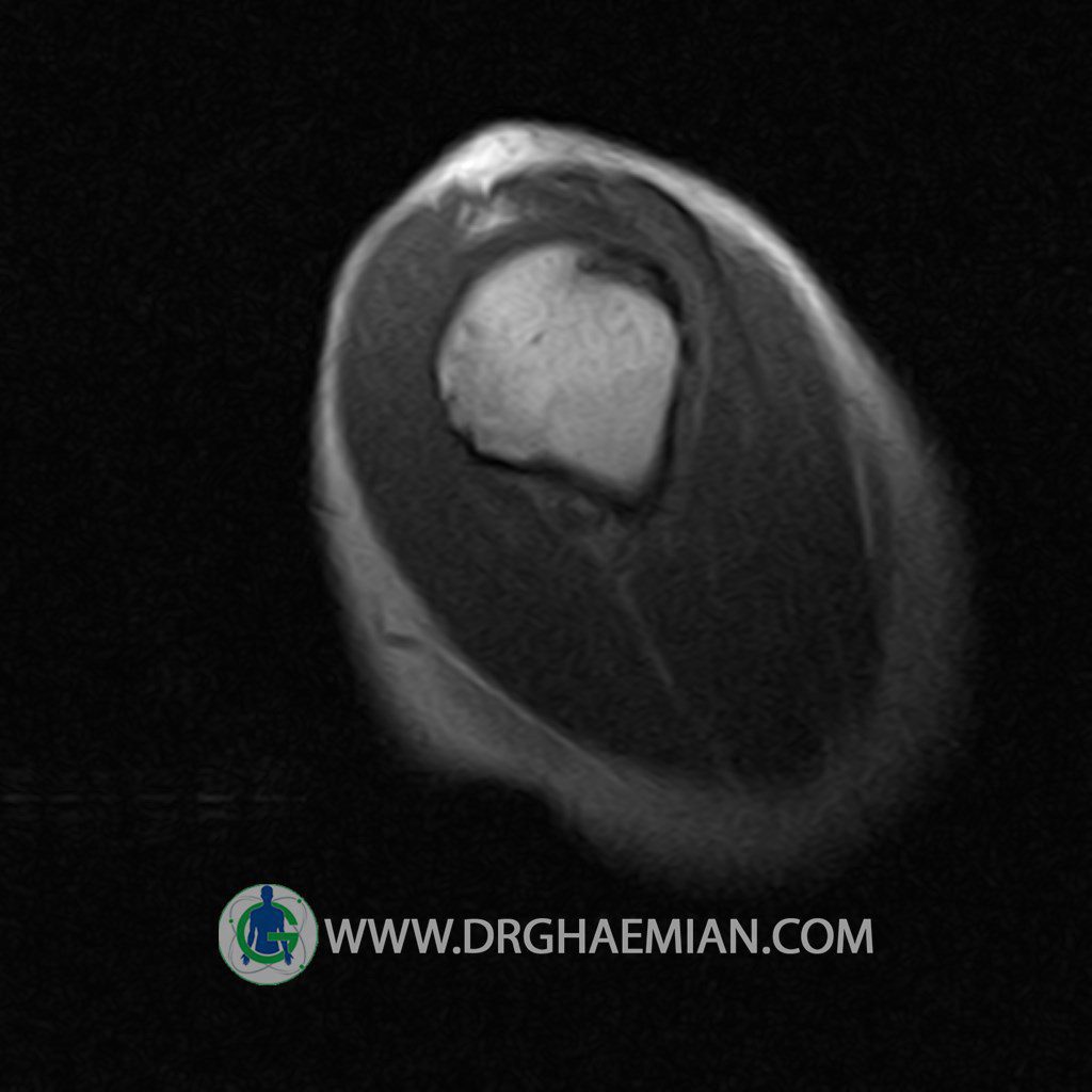
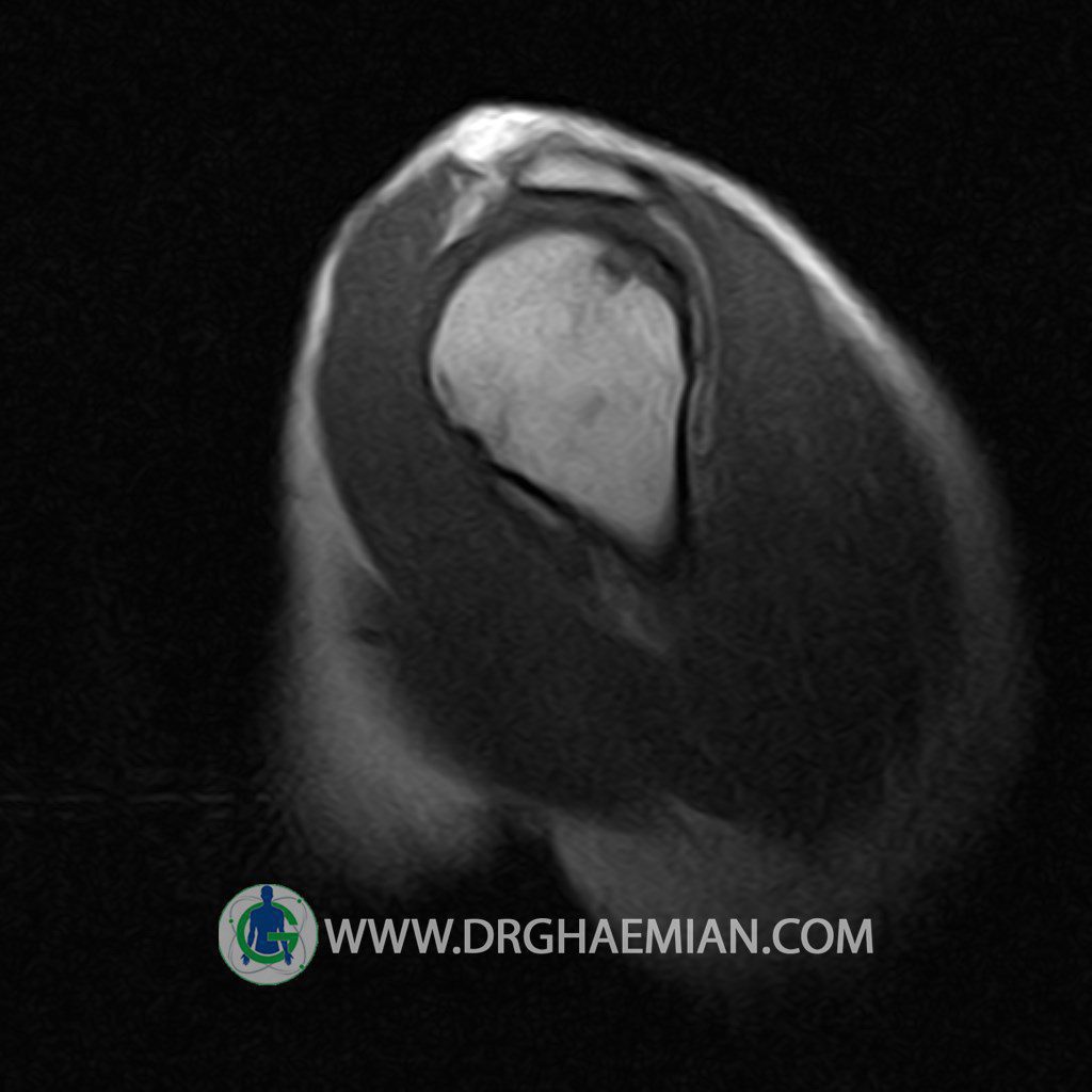
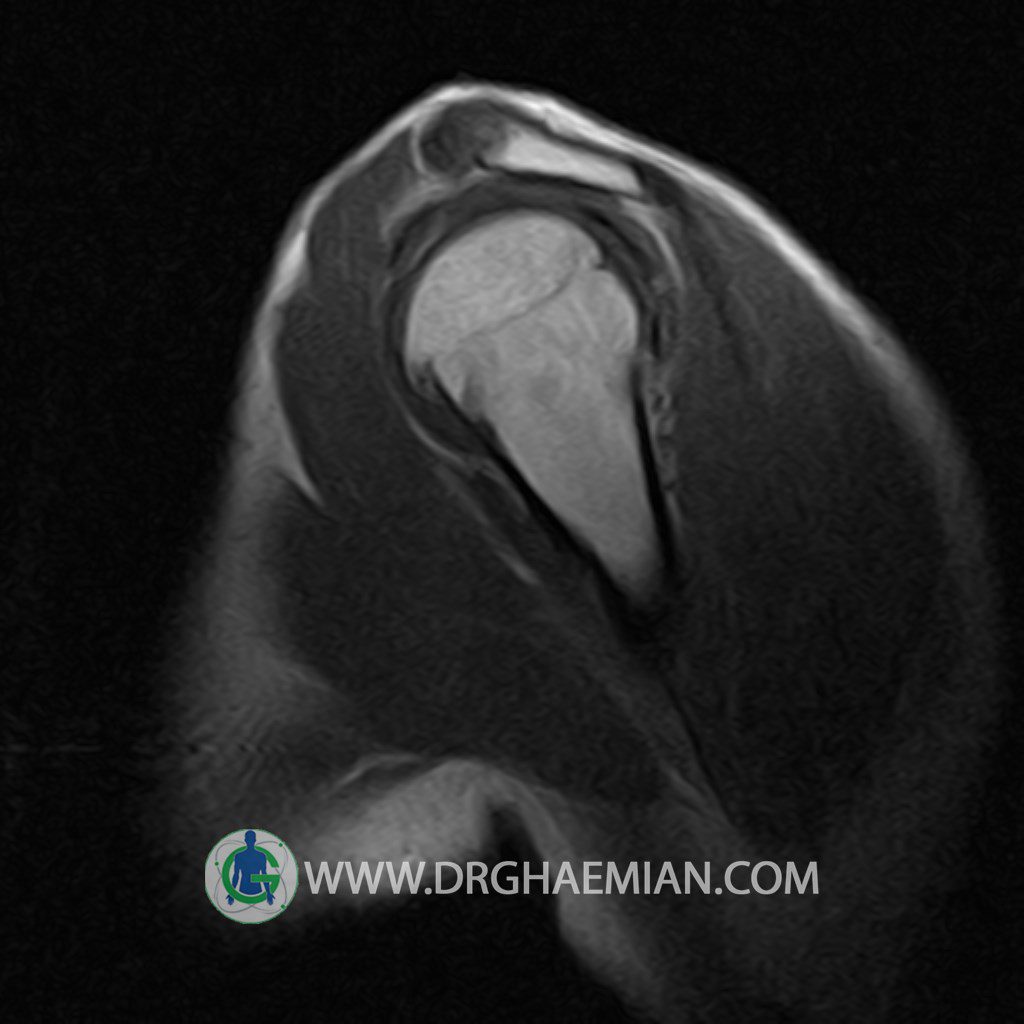
RIGHT SHOULDER MRI
(Without contrast)
Technique: Axial T1 and GE , coronal T1 , T2 , sagittal T1 , T2 .
REPORT:
The humeral head has normal configuration and articulates properly and parallelism with the normally developed glenoid .
The articular surfaces are smooth and show normal cortical thickness .
The width of the joint space is normal .
The glenoid labrum is intact on all sides .
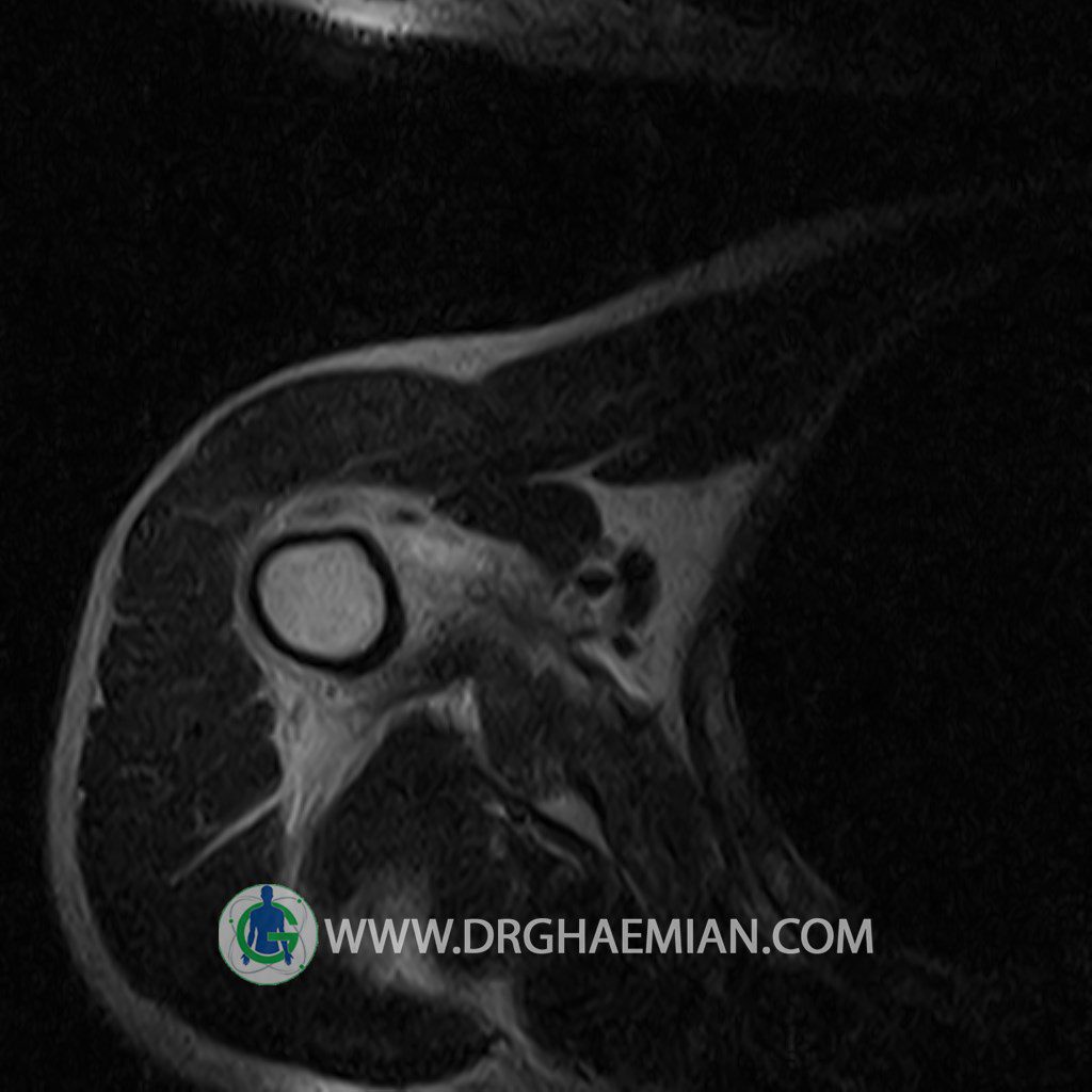
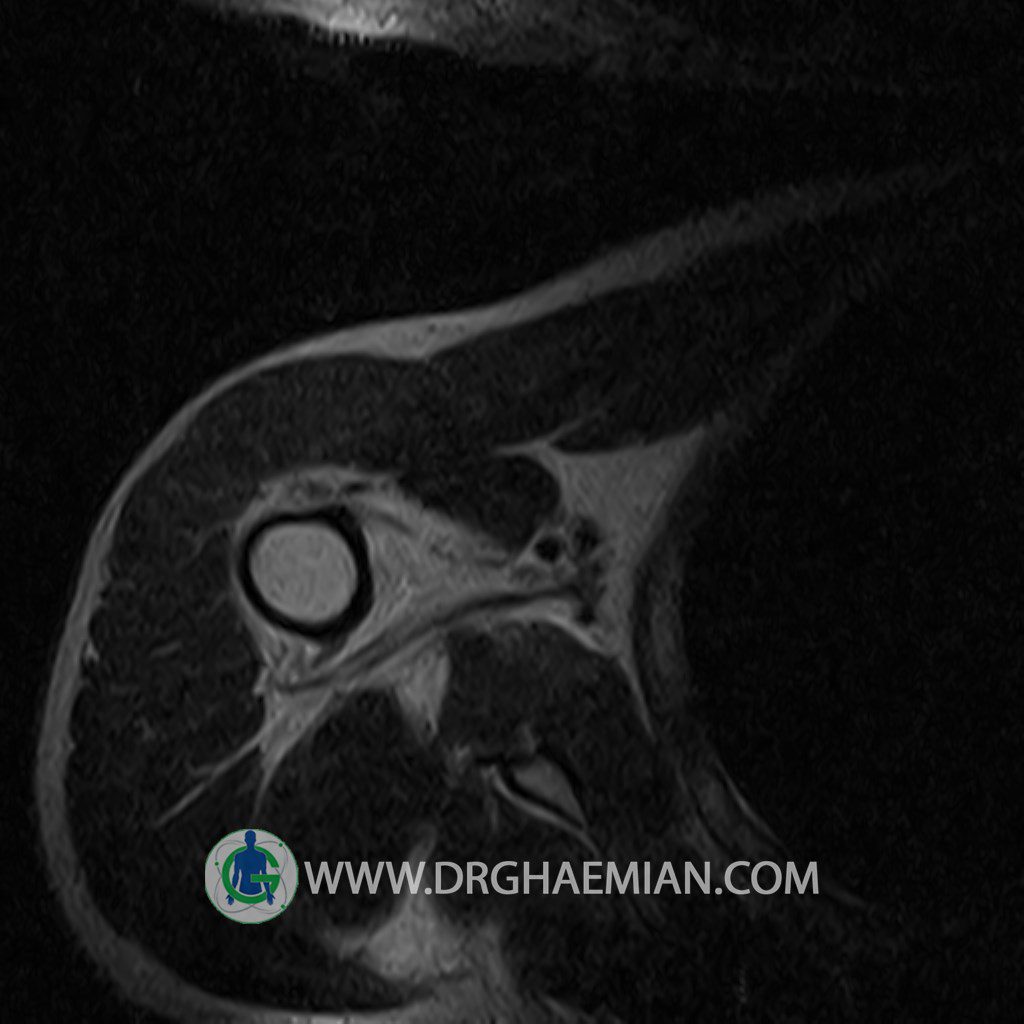
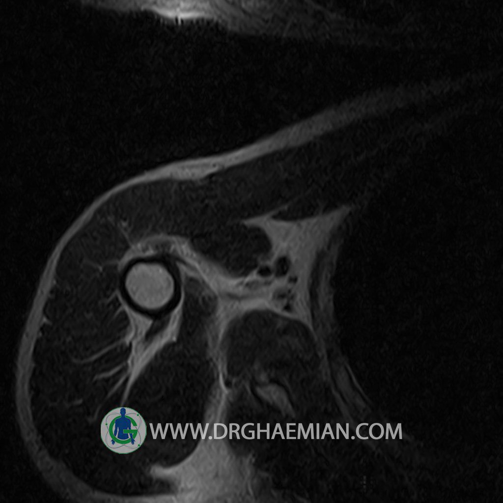
The biceps tendon appears normal and occupies a normal position in the bicipital groove.
The other muscles that cover the shoulder joint appear normal , as do imaged portions of the lungs and soft tissues .
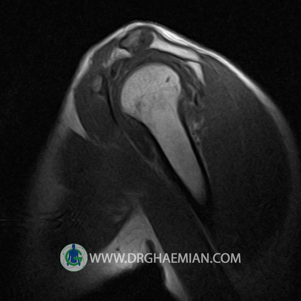
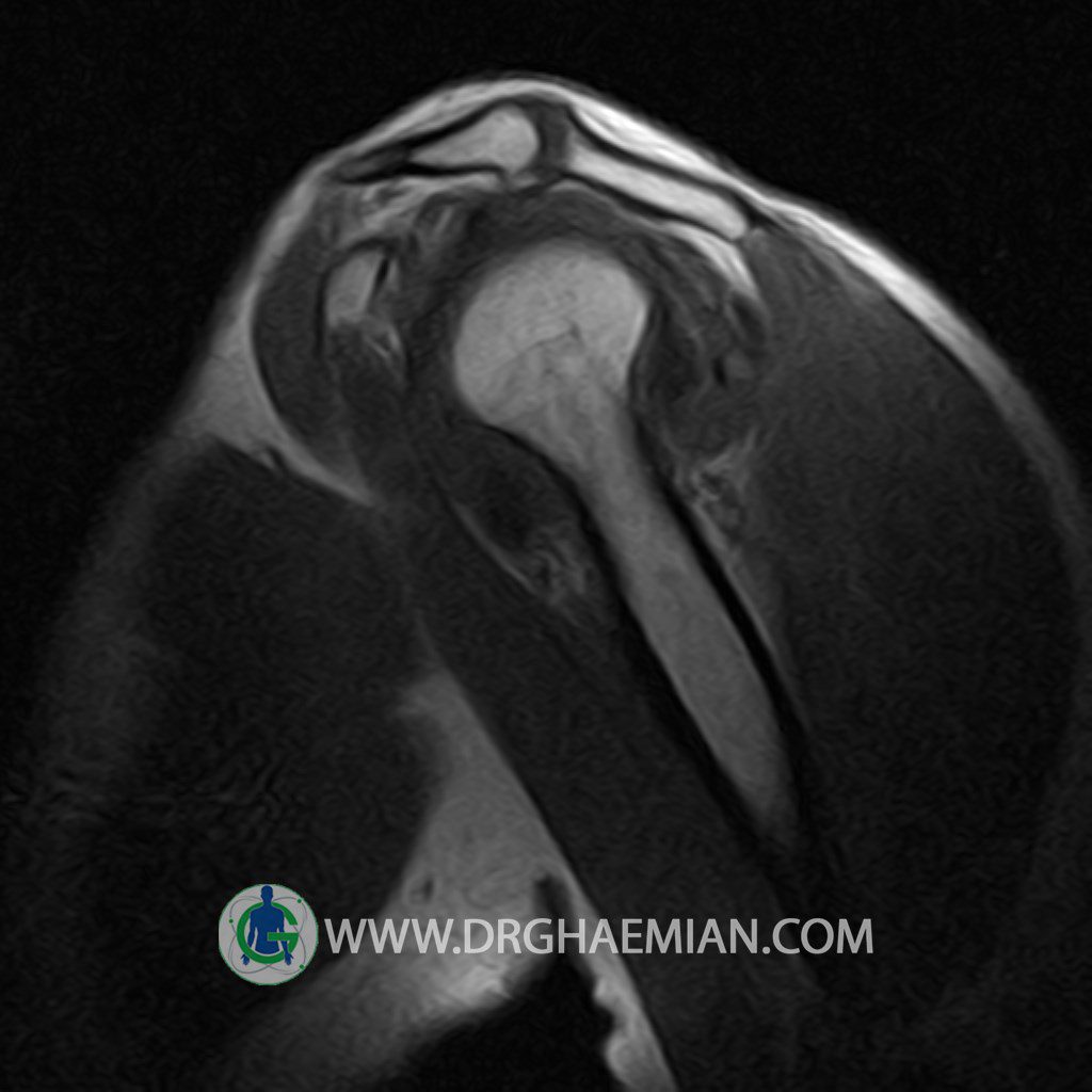
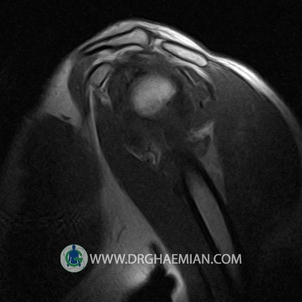
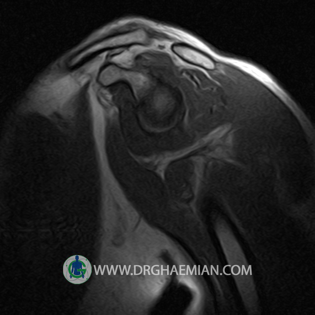
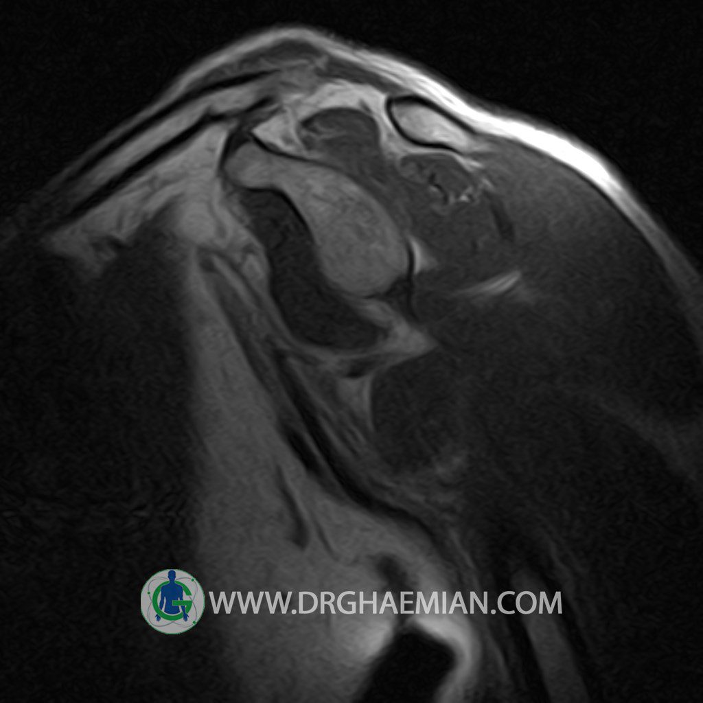
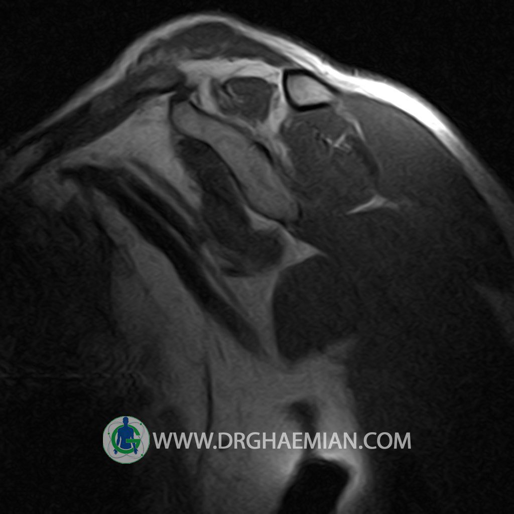
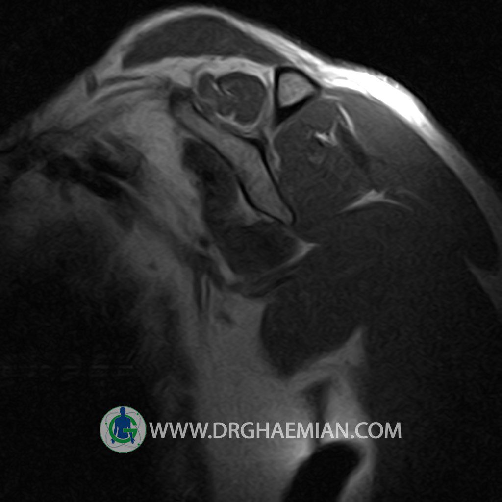
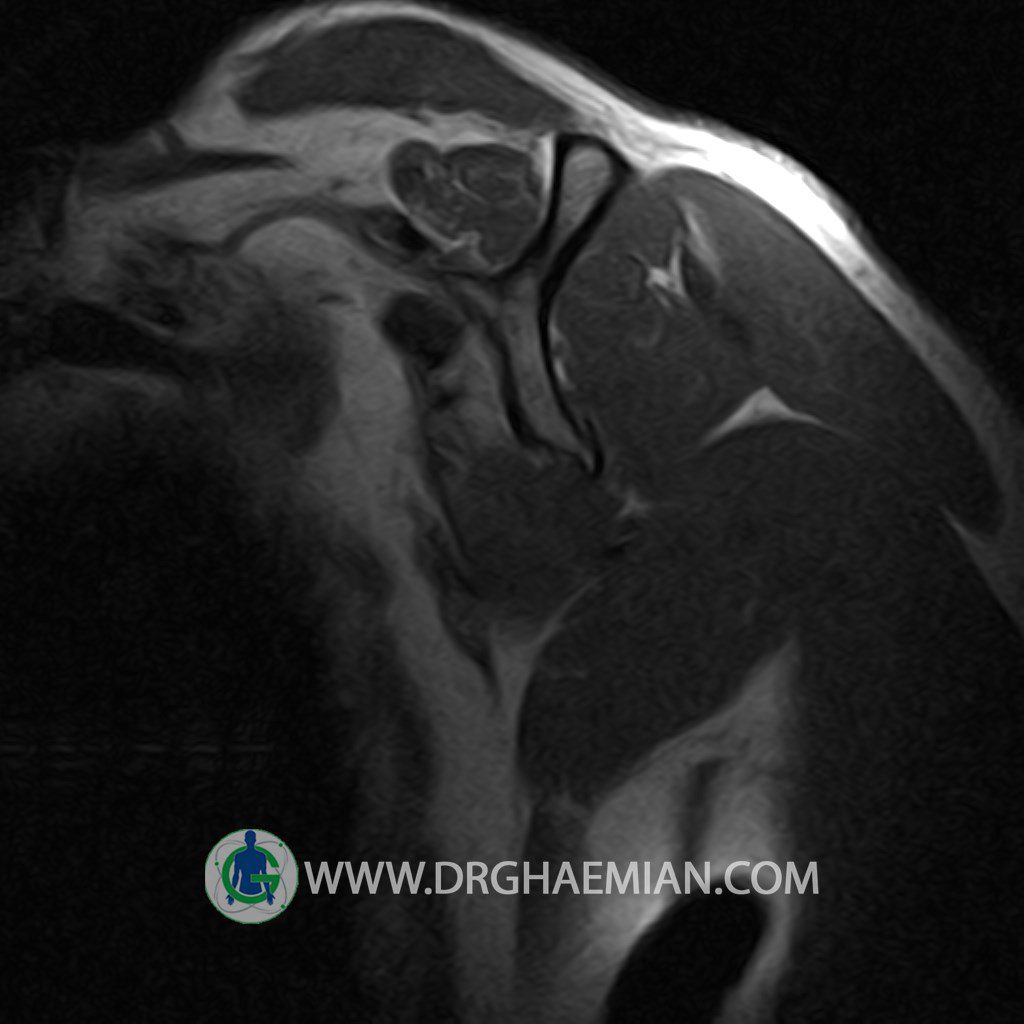
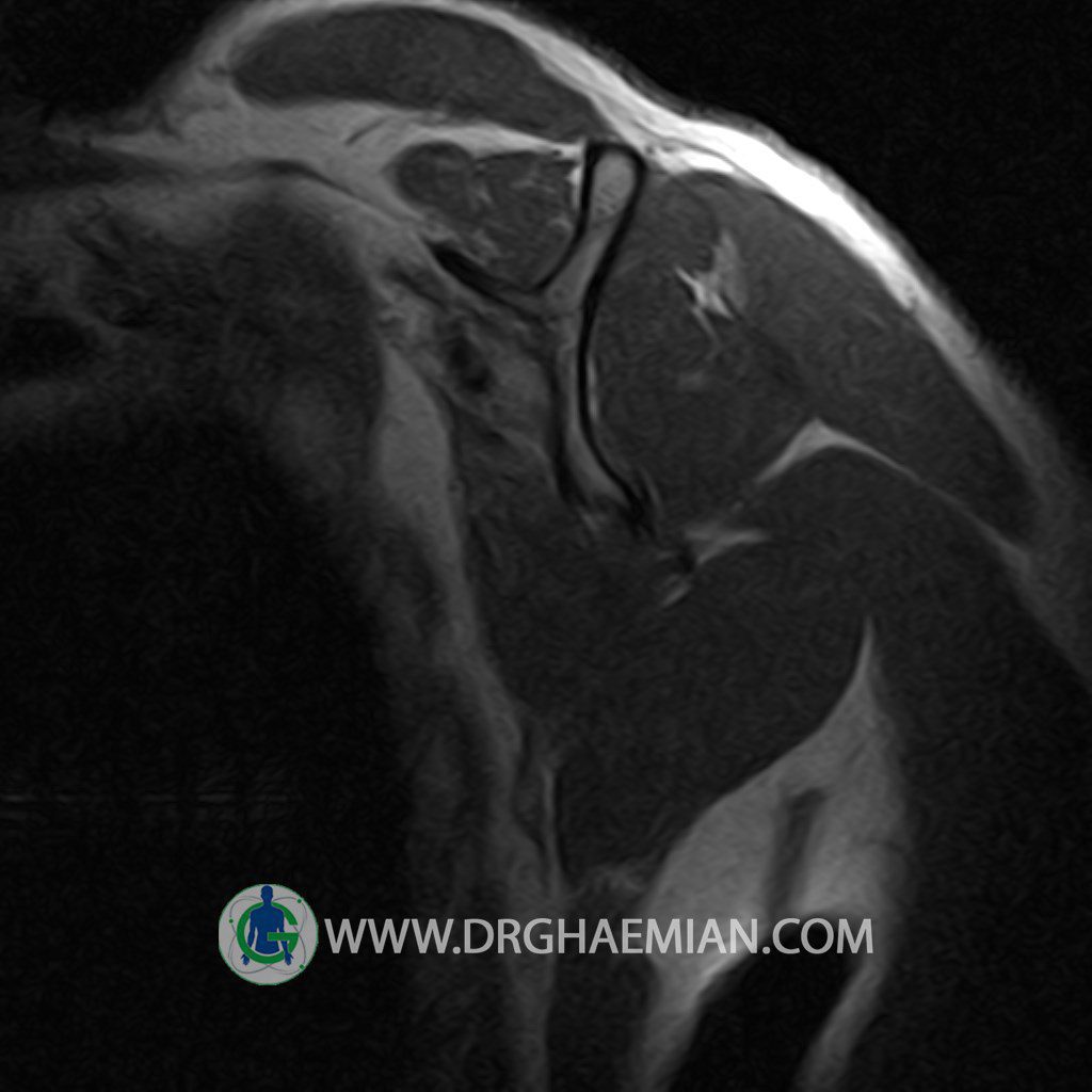
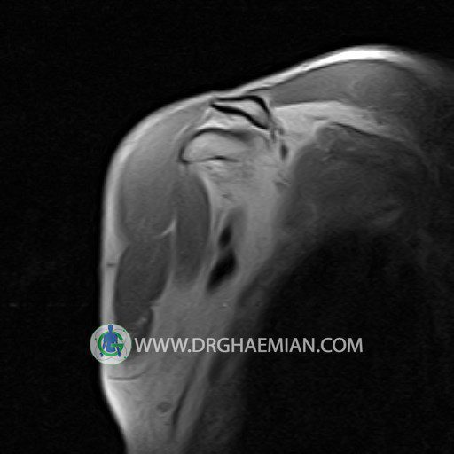
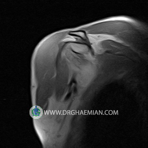
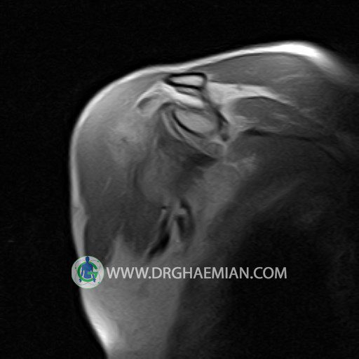
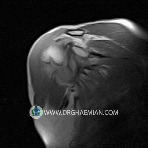
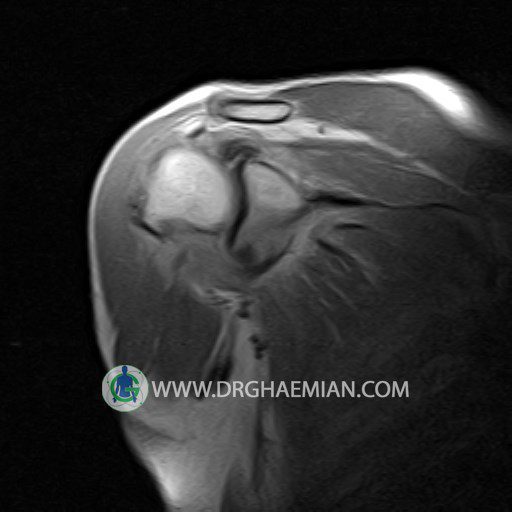
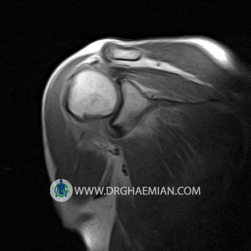
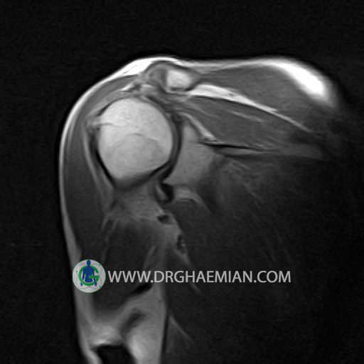
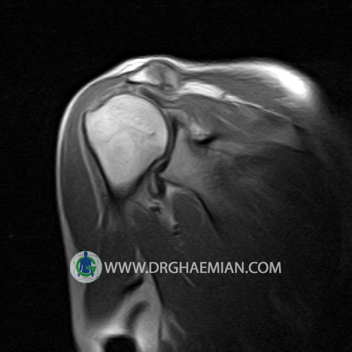
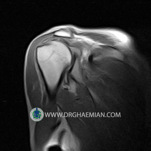
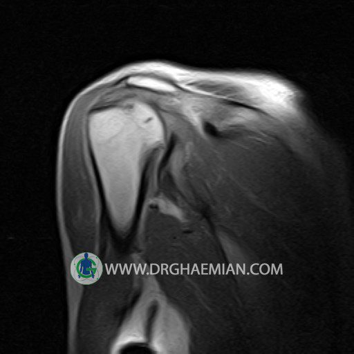
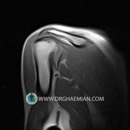
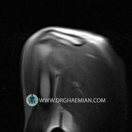
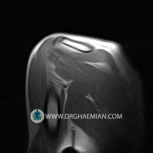
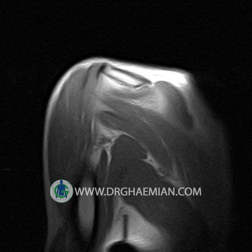
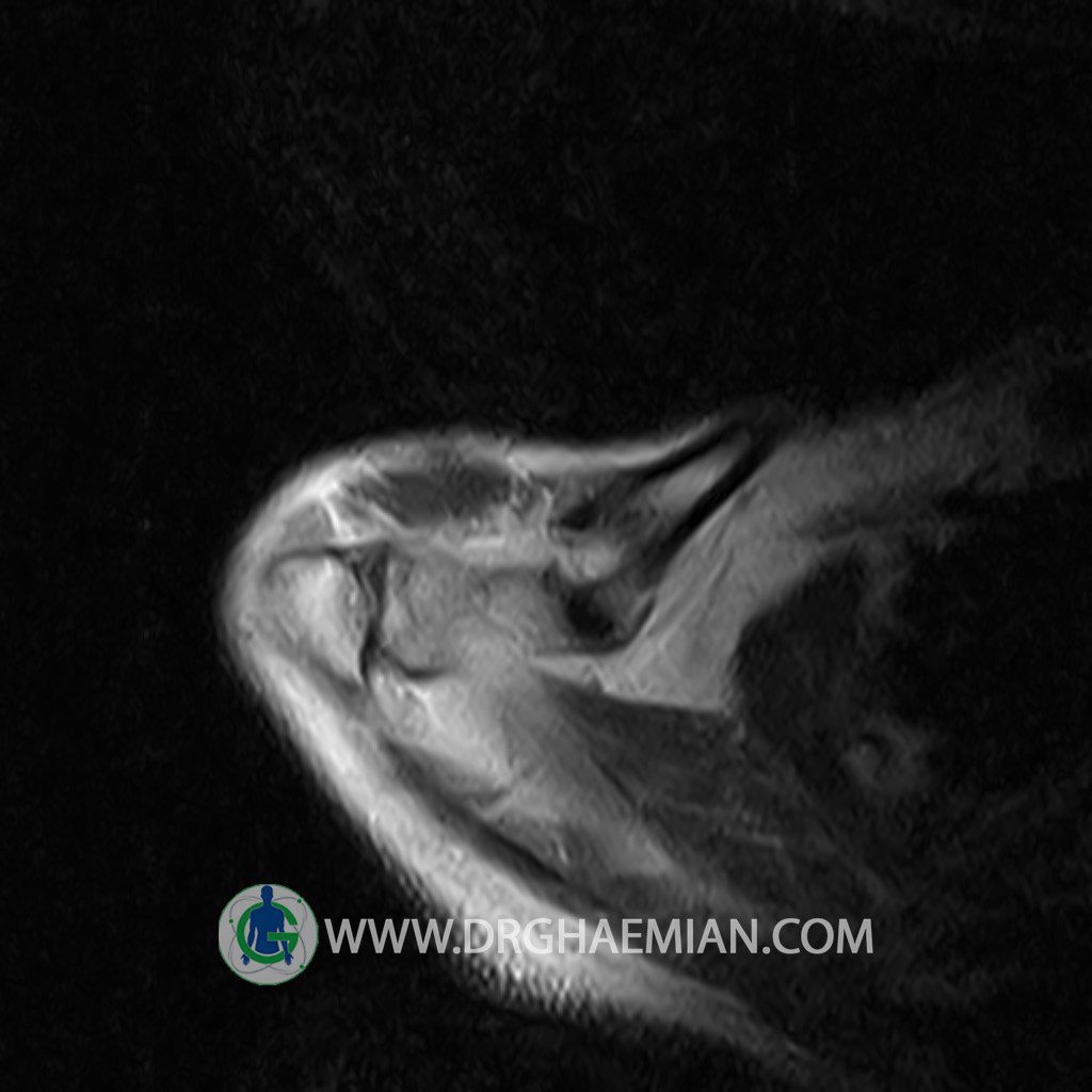
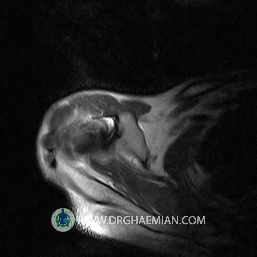
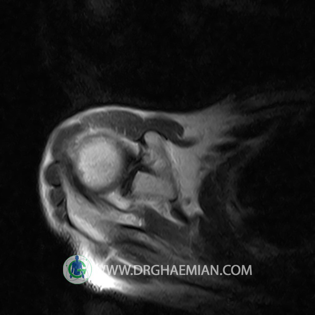
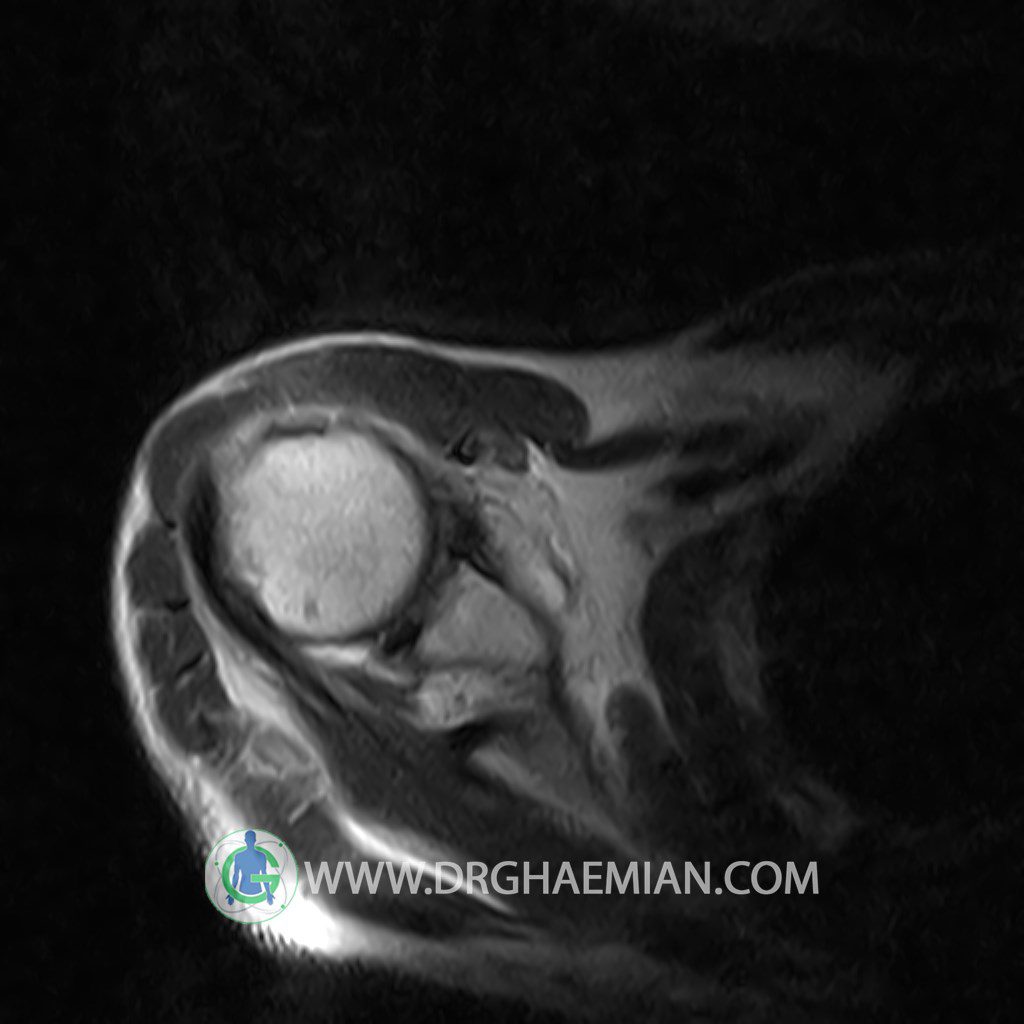
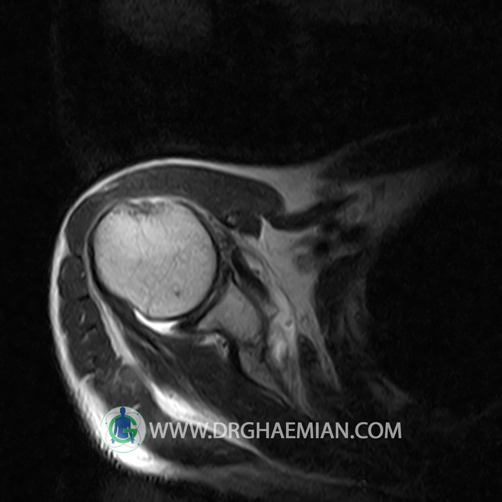
– A – C joint hypertrophy & synovitis with subacromial – subdeltoid bursitis
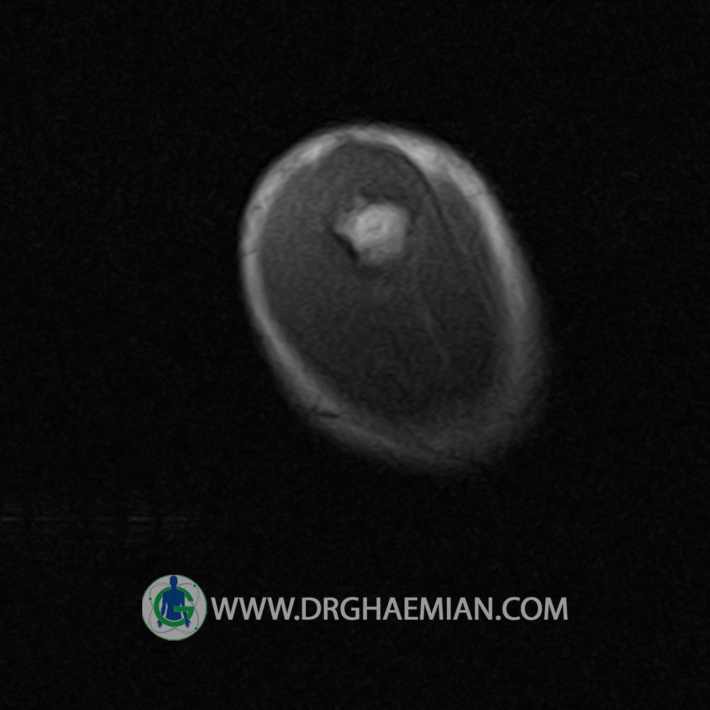
– Full thickness partial with tearing in anterior of supra – spinatus tendon with focal bone bruise in greater tuberosity of humerus
– Subscapularis & Infraspinatus tendon sprain
– Glenohumeral joint effusion
are seen
