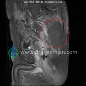
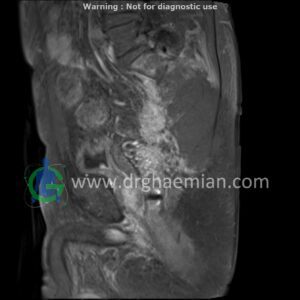
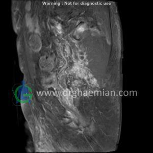
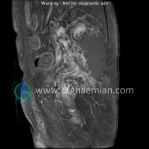
ام آر آی لگن یک روش تصویربرداری است که از طریق دستگاهی با آهنرباهای قوی و امواج رادیویی از ناحیه بین استخوان های ران تصاویری می سازد. این قسمت از بدن را ناحیه لگنی می گویند. در این بیمار توده نئوپلاسی در ساکروم و متازتاز استخوانی دیده میشود.

گزارش پزشک
SACROILIAC JOINTS MRI ( with and without contrast )
Technique : Axial , coronal T1 , Axial , coronal T2 , coronal T2 fat sat Axial T1 post Gd .
REPORT :
The sacroiliac joints are normally shaped with normal development of the sacrum and iliac wings and a normal – appearing lumbosacral junction .
The joint space is of normal width on both sides and have smooth , sharply defined contours .
The sacrum and iliac wings also contain normal bone marrow and present smooth and intact cortical boundaries .
The sacral neuroforamina are of normal width and contours .
The nerve filaments shows a normal course and diameter , and the width of the sacral spinal canal is normal .
tumoral infiltration in sacrum ( 60x110x130mm ) with bone destruction , soft tissue component , invasion to rectum , post contrast enhancement , suggestive for neoplasic lesion & bone metastasis
are seen
.COMMENT : image guided CNB is recommended
