پزشکان اغلب از تصویربرداری ام آر آی برای تشخیص و درمان عارضه های پزشکی که فقط با استفاده از اشعه ایکس یا میدان مغناطیسی و امواج رادیویی قابل مشاهده است، استفاده می کنند. دستگاه ام آر آی تصاویر دقیق از ساختار های داخلی بدن ایجاد می کند. در این کیس استئومیلیت لگن، آتروز و سنیویت دیده می شود.
گزارش پزشک :
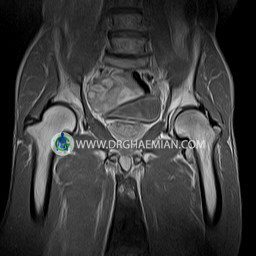
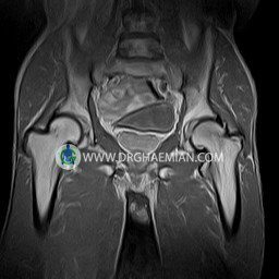
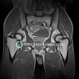
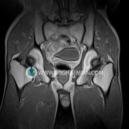
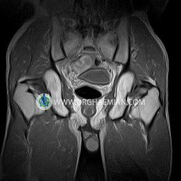
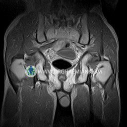
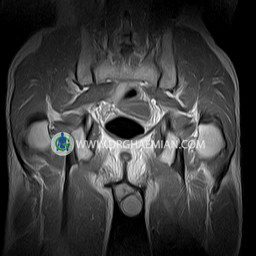
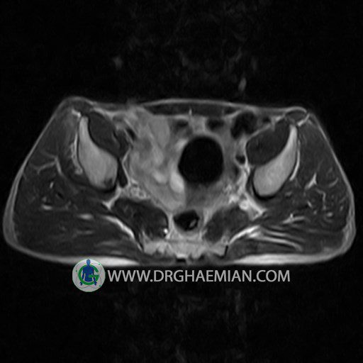
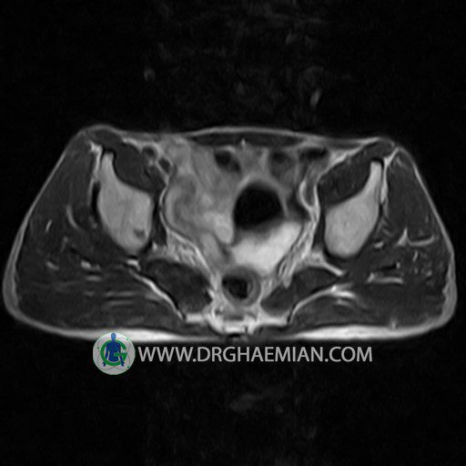
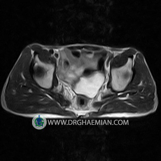
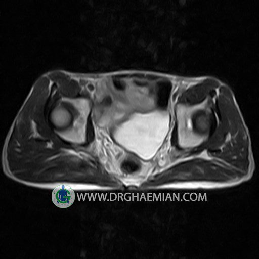
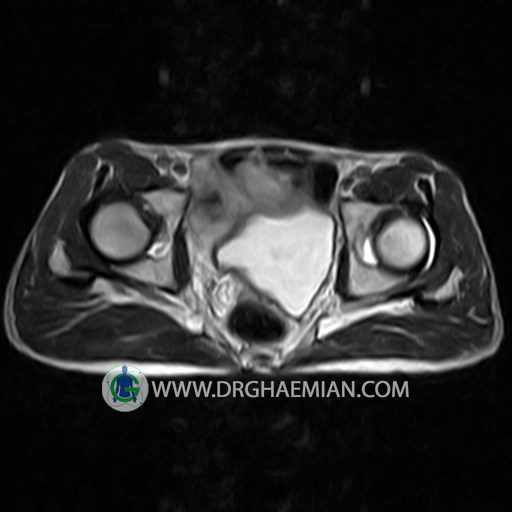
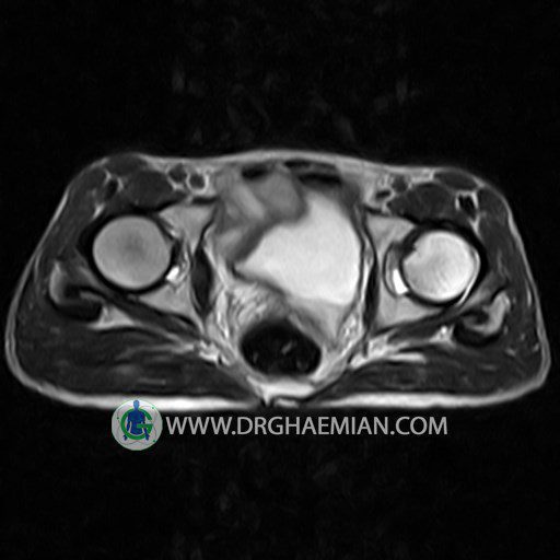
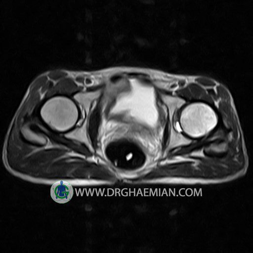
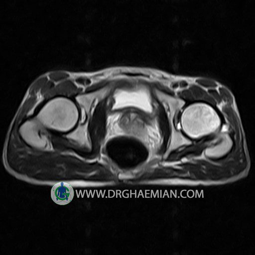
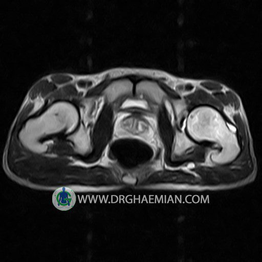
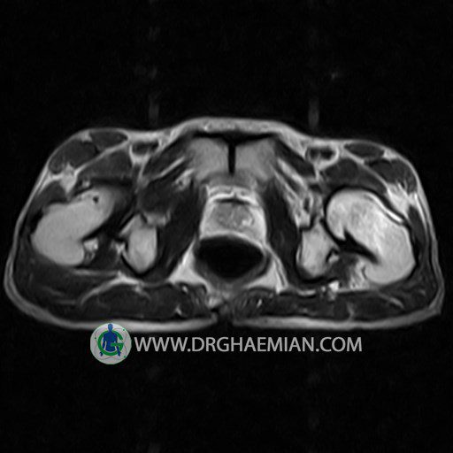
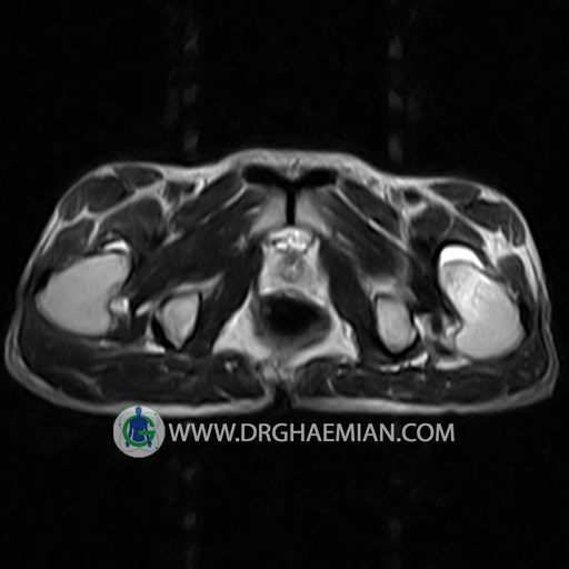
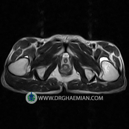
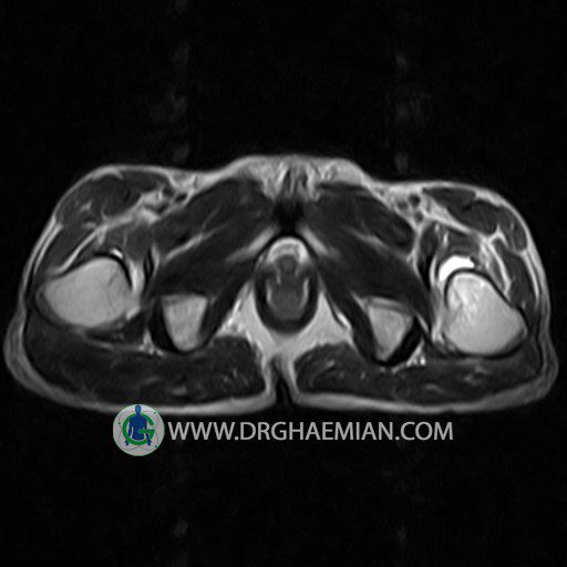
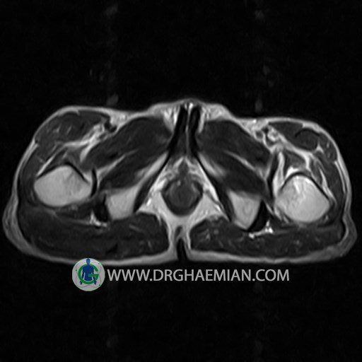
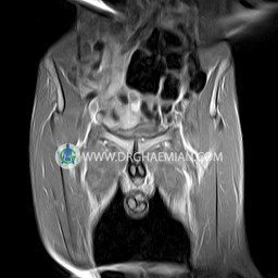
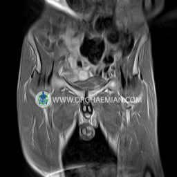
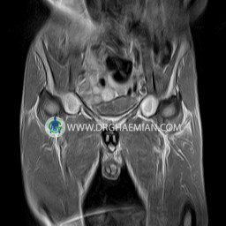
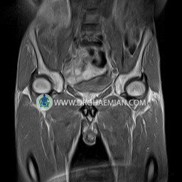
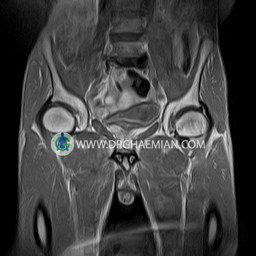
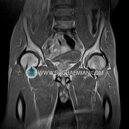
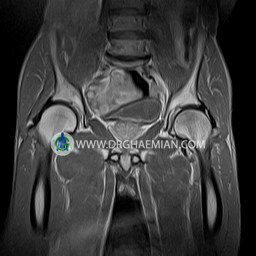
HIP JOINT MRI
( without contrast )
Technique : coronal STIR , coronal T2 , Axial T1 , axial T2 .
REPORT:
The femoral heads and acetabula are normal shape , signal intensity and the femoral heads are well covered by the acetabular margins .
The joint spaces are of normal width without fluid collection .
The articular surfaces are smooth and congruent and show normal cortical thickness .
Each femoral shaft has normal margins and contains a normal bone marrow signal .
The imaged muscles and the lesser pelvis show no abnormalities .
– Heterogeneous signal change (high T2/STIR , low T1) in proximal metaepiphysis of left femur without articular surface irregularity suggestive for bone bruise (stress fracture?), osteomyelitis & arthritis and marrow infiltration (less probable)
– Left hip joint effusion suggestive for synovitis
are seen.
COMMENT: Clinical correlation and MRI with contrast are recommended.
