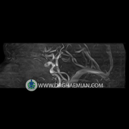
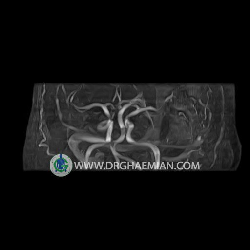
ام آر آنژیوگرافی با استفاده از آهنربایی قوی، امواج رادیویی و کامپیوتری هوشمند عروق خونی را ارزیابی کرده و به یافتن موارد غیرعادی کمک میکند. این تصویربرداری از تششعات استفاده نمی کند و ممکن است از کنتراست (ماده حاجب) استفاده شود. در این کیس ترومبوز فوکال بیمار مشاهده می شود.


گزارش پزشک :
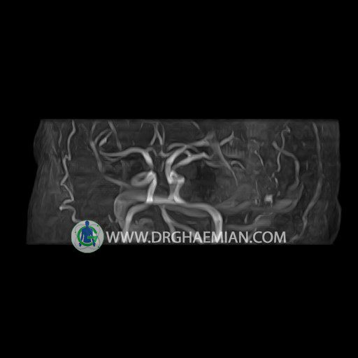
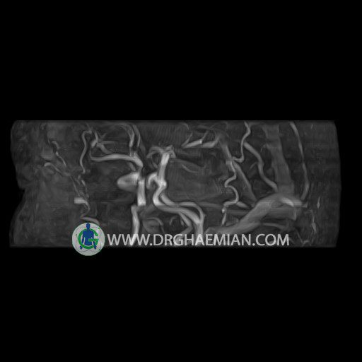
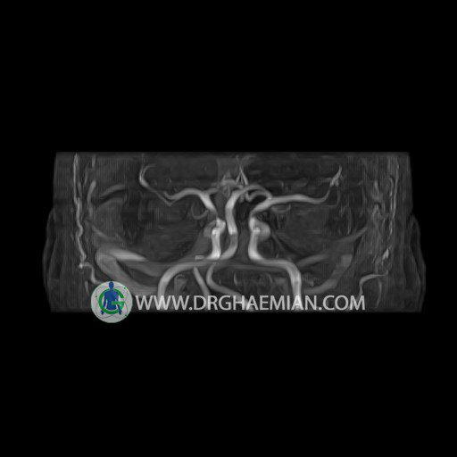
CEREBRAL MRA
REPORT :
The internal carotid arteries show normal course , caliber and are symmetrically disposed.
Each carotid siphon is normal , showing no displacement or extrinsic compression and intraluminal signal intensity is homogeneous.
The middle cerebral artery arises normally from the internal carotid on each side and forms normal insular loops shows homogeneous signal intensity lumen with no circumscribed vascular narrowing or dilatation.
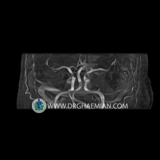
The anterior cerebral artery shows no signs of narrowing or displacement.
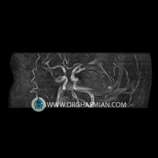
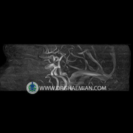
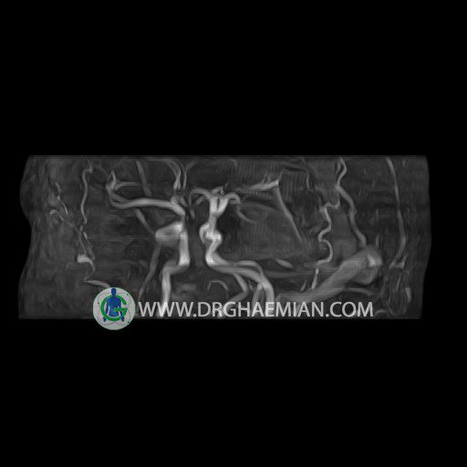
The basilar artery shows a normal course ,caliber and divides into
normal posterior cerebral arteries.
The anterior and posterior communicating arteries on each side are normally developed and of normal caliber.
No segments show convolution or circumscribed dilatation.
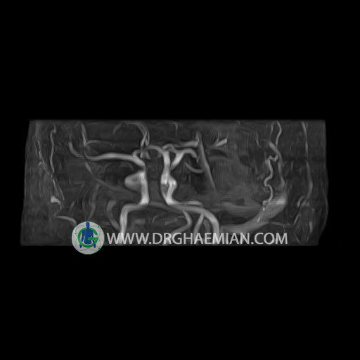
The other evaluable portions of the neurocranium show no abnormalities.
– Focal filling deffct in left transverse sinus suggestive for focal thrombosis
is seen
