سی تی اسکن شکم و لگن یکی از روش های تصویربرداری با سی تی اسکن است. این روش با استفاده از اشعه ایکس تصاویر عرضی از ناحیه شکمی و لگنی ایجاد میکند.در این کیس همانژیوم کبدی و… دیده میشود

گزارش پزشک
Spiral CT Scan of Abdomen & Pelvis With & Without Contrast (3phasic Study)
384-slice CT scan System
Dear colleague,
Non-contrasted and thereafter, contrast-enhanced abdominopelvic multislice spiral CT (MSCT) scan was performed.
On obtained slices, the findings are as follow:
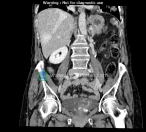
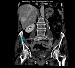
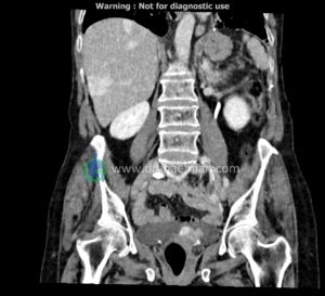
There are at least five foci of early arterial enhancing lesions, some of them with peripheral nodular discontinues enhancement and the rest on left lobe, shows homogenous flash-like enhancement, associated with progressive centripetal filling on porto-venous & delay images with also evidence of AP portal shunts.
All findings are mostly compatible with hemangioma, maximum size is located in subscapular side of segment V in right liver lobe measuring 25×36mm and in left lobe segment IVA measured 31×34mm.
Wedge shaped low attenuated changes is seen in anterior aspect of right kidney -inferior pole, seems to be as sequel of cortical infarct.
Mild atherosclerotic changes are detected in infra-renal segment of abdominal aorta.
There is evidence of cystic appearing lesion in right adnexa, measuring 31×42mm, with no evidence of apparent enhancing mural nodule, however, correlation with sonography is requested.
Intramural myoma is seen in posterior wall of uterine fundus (8.5mm, Figo:4)
Osteoporotic changes is detected in lumbosacral spine and pelvic girdle.
The pancreas and spleen are of normal appearance and homogeneous attenuation value, lacking any space-occupying lesion.
Intra- and extrahepatic bile ducts are of normal caliber.
The gallbladder is of normal volume and lacks any CT-detectable abnormal finding.
The adrenal glands are normally seen, bilaterally.
Kidneys are of normal dimensions and attenuation value, revealing ordinary excretory function, bilaterally, lacking any solid lesion, intrarenal stone or hydronephrosis, on either side.
The ureters and bladder are normally seen.
Detectable portions of stomach and bowel loops, as well as abdominopelvic vascular structures are ordinarily depicted, considering patient’s age.
Neither significant abdominopelvic lymphadenopathy, nor ascites is found.
There is no pleural effusion, on either side.
