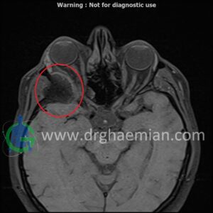
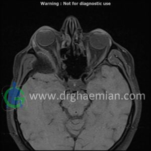
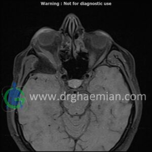
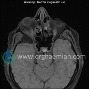
ام آر آی اوربیت با استفاده از آهنربا و امواج رادیویی تصاویری از اوربیت ها، اعصاب، عضلات و بافت های اطراف آن ایجاد می کند. در این کیس مننژیوم مغزی در پشت چشم بیمار دیده میشود.

گزارش پزشک
ORBIT MRI
(with and without contrast)
Technique : Axial T1 , Axial , sagittal , coronal FSE T2 , coronal T1, sagittal fat sat T2 , Axial , sagittal T1 post Gd .
REPORT :
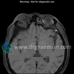
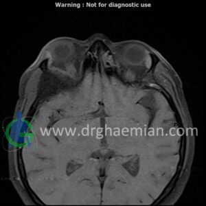
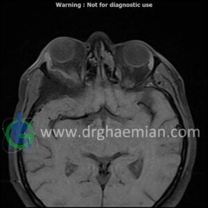
No foci of bone destruction , no circumscribed expansion of the bony or soft – tissue components of the orbital are evident .
The globes are symmetrical and of normal size and the ocular contents show normal signal characteristics .
The optic nerve has normal course and caliber on each side .
The retrobulbar fat, ophthalmic vein and lacrimal apparatus are unremarkable .
Evaluable portions of the neurocranium and paranasal sinuses show no abnormalities .
thickening of posterolateral of right orbit wall ( low T1/T2/STIR ) with mass effect on right temporal pole & right orbital content/proptosis , with adjacent dural thickening & post contrast enhancement ( 33x41mm – without significant change to prior MRI – 1403/09/20) suggestive for :
1.meningioma
2.fibrosis plasia with dural thickening & less probably sclerotic bone metastasis
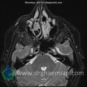
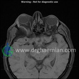
mucosal thickening in paranasal sinuses
retention cyst in left maxillary sinus
concha bullosa in right & left middle concha
are seen
