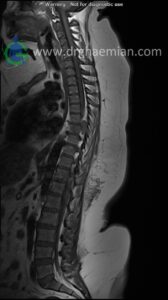
ام آر آی کمر از طریق انرژی آهنربا های قوی تصاویری از قسمت پایین ستون فقرات ( گودی کمر) ایجاد می کند. در این کیس بیمار با تومور گانگلیون نوروم…. دیده میشود
گزارش پزشک
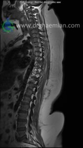
THORACIC SPINE MRI
(Without contrast with sagittal total spine reconstruction)
Siemens MRI ( magnetom altea 1.5 tesla )
Technique : Sagittal T1 , T2 , Axial T2, Sagittal & Coronal myelogram. REPORT:
The thoracic spine shows a smooth kyphotic curvature with normal alignment. The vertebral bodies and endplates are normal in shape and signal intensity. The intervertebral disk spaces are normal height . The bony spinal canal has normal width. The imaged soft tissues show no abnormalities . Paravertebral stripe is normal in shape and signal intensity .
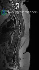
– Thoracic discs dehydration
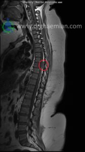
– A well defined intradural extramedullary elongated mass lesion (8×25×30mm) at level of T6/T7 with mass effect &
anterior displacement of thoracic cord with extension to left neural foramina suggestive for neurogenic tumor such as
ganglion neuroma
are seen.
COMMENT: MRI with contrast and tissue diagnosis are recommended
