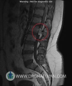
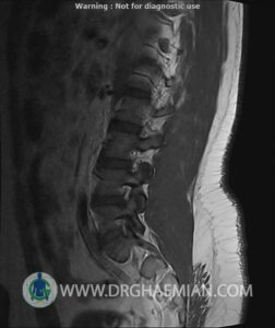
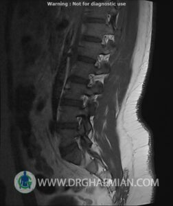
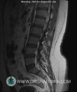
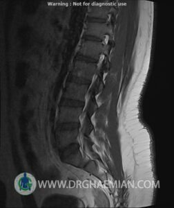
ام آر آی کمر از طریق انرژی آهنربا های قوی تصاویری از قسمت پایین ستون فقرات ( گودی کمر) ایجاد می کند. در این کیس تومور نخاعی تومور اپاندیموم به ابعاد 11*25 میلی متر و از بین رفتن کامل فضای CSF و … دیده میشود
گزارش پزشک
LUMBOSACRAL SPINE MRI
(With contrast)
Siemens MRI ( magnetom altea 1.5 tesla )
.Technique : Pre contrast coronal T1 fat sat , post contrast Axial coronal T1 fat sat and sagittal T1
:REPORT
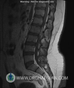
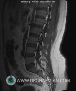
The lumbar spine shows a smooth lordotic curvature with normal alignment
The vertebral bodies and endplates are normal in shape and signal intensity
The intervertebral disk spaces are normal height and signal intensity
The bony spinal canal has normal width which , neural foramina and posterior element displays normal nature
The imaged soft tissues show no abnormalities
The conus medullaris terminates normally at L1
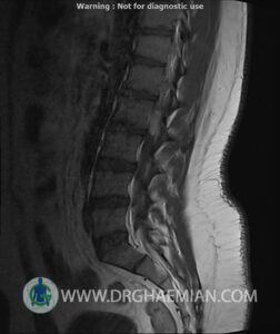
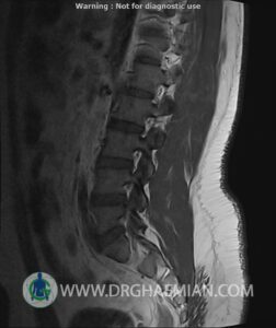
Paravertebral stripe is normal in shape and signal intensity -An intrathecal mass at the lower L2 level shows intermediate T1 & heterogeneous increased T2 signal and vividly enhancement
It displaces the cauda equina posteriorly with near complete effacement of CSF space and inseparable from the filum terminale which runs along its posterior border
. No evidence of osseous remodeling
. No mass or abnormal enhancement elsewhere within the spine
:CONCLUSION
.Intradural extramedullary mass lesion (11x25mm) suggestive for myxopapillary ependymoma
