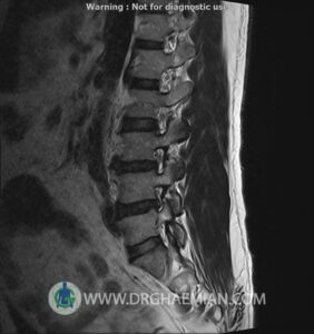
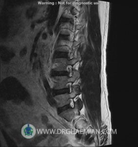
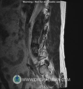
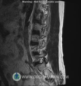
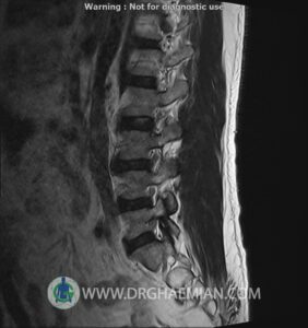
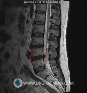
ام آر آی کمر از طریق انرژی آهنربا های قوی تصاویری از قسمت پایین ستون فقرات ( گودی کمر) ایجاد می کند. در این کیس بیرون زدگی مهر های کمری L5/L4 و … دیده میشود
گزارش پزشک
LUMBOSACRAL SPINE MRI
(Without contrast)
Siemens MRI ( magnetom altea 1.5 tesla )
Technique : Sagittal T1 , T2 , Axial T2, Sagittal & Coronal myelogram
:REPORT
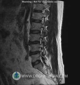
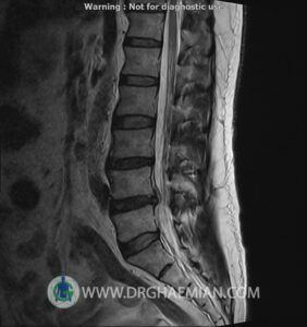
.The lumbar spine shows a smooth lordotic curvature with normal alignment
.The imaged soft tissues show no abnormalities
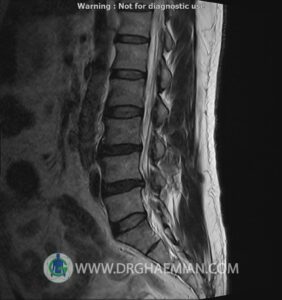
.The visualized cord and filum terminalis are normal
. The conus medullaris terminates normally at L1
.Paravertebral stripe is normal in shape and signal intensity
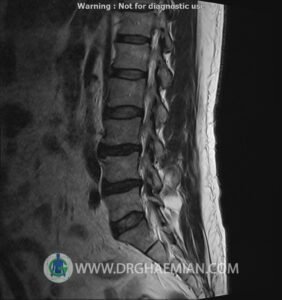
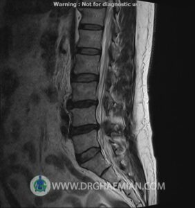
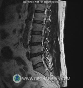
– L3/L4 spondylosis, disc space narrowing, dehydration and bulging
– L4/L5 retrolisthesis grade 1, disc dehydration, bulging, right central protrusion with thecal compression and canal stenosis
are seen
