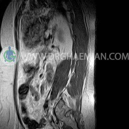
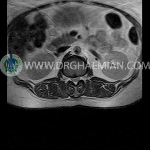
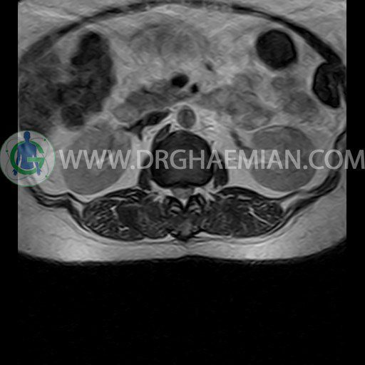
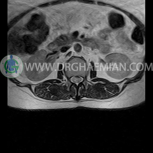
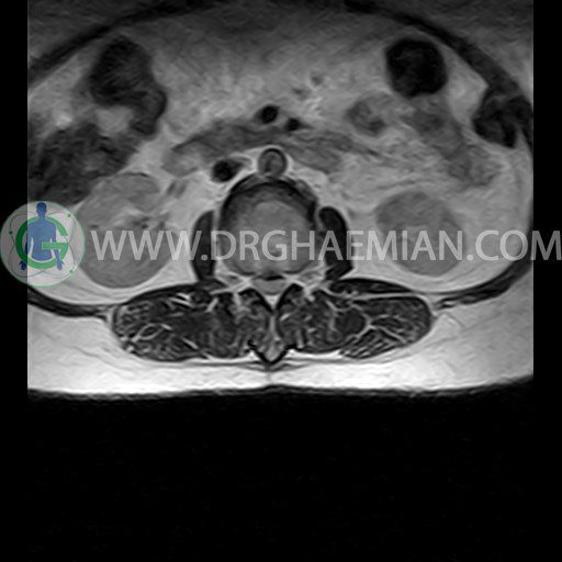
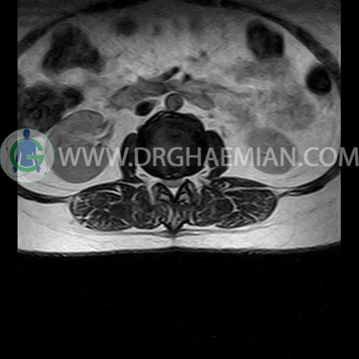
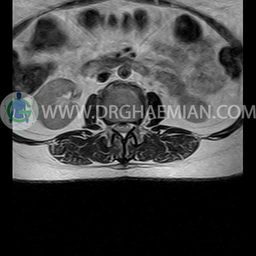
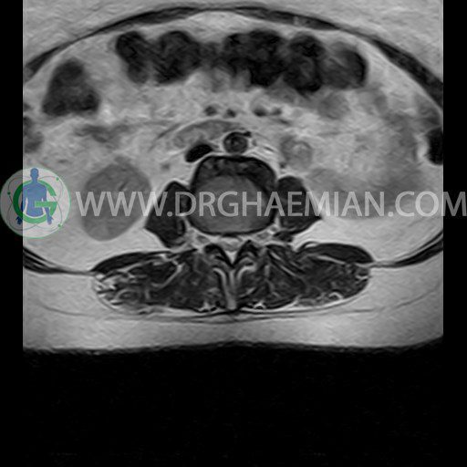
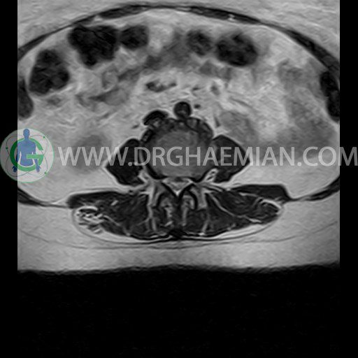
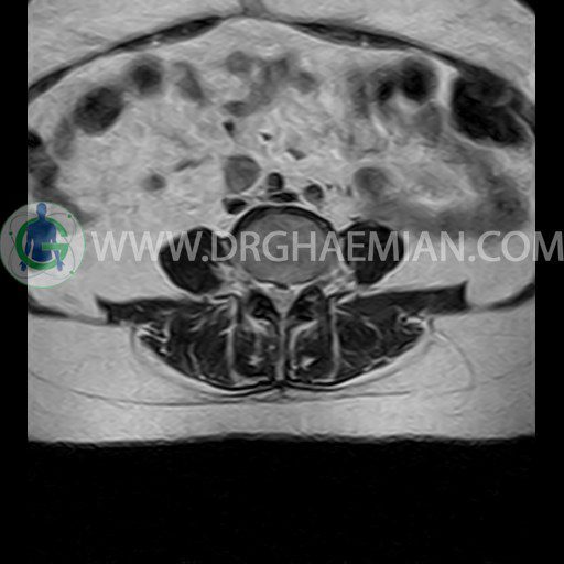
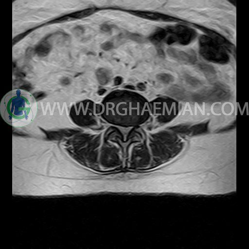
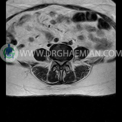
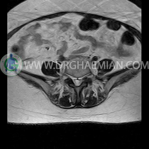
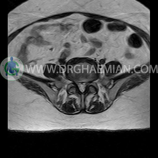
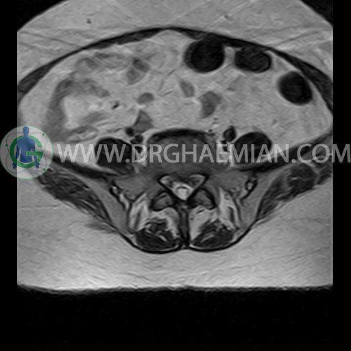
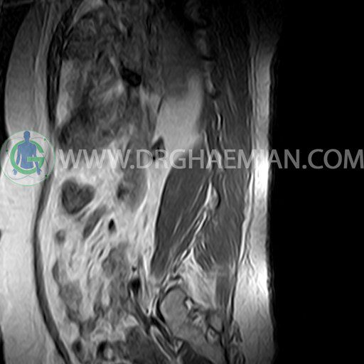
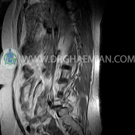
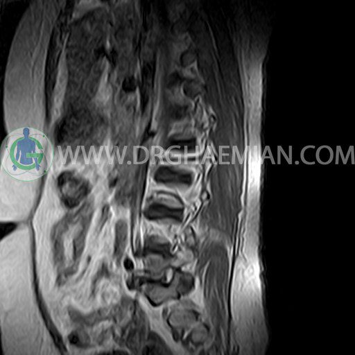
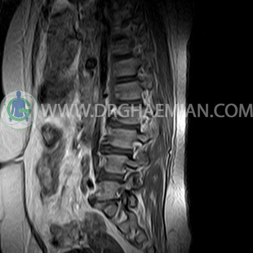
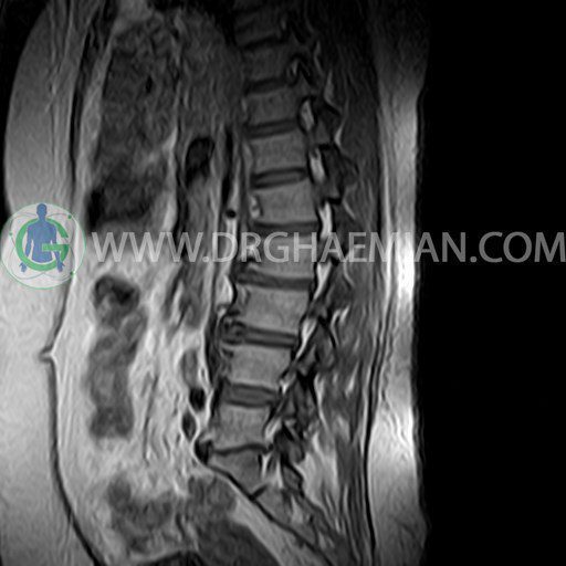
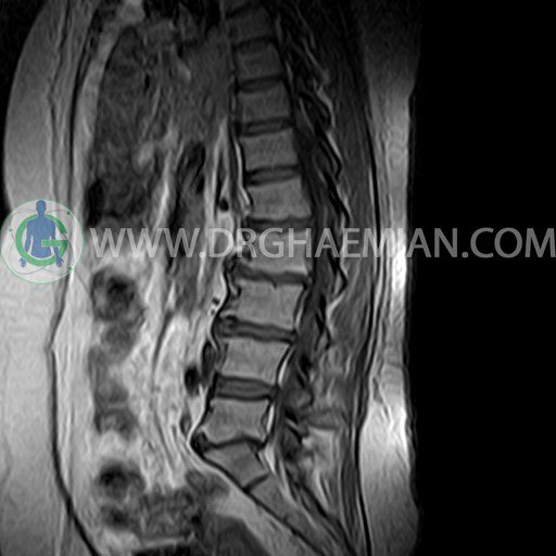
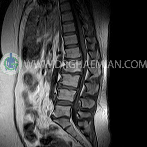
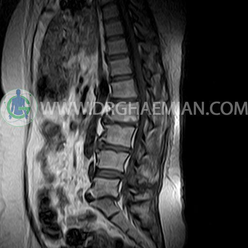
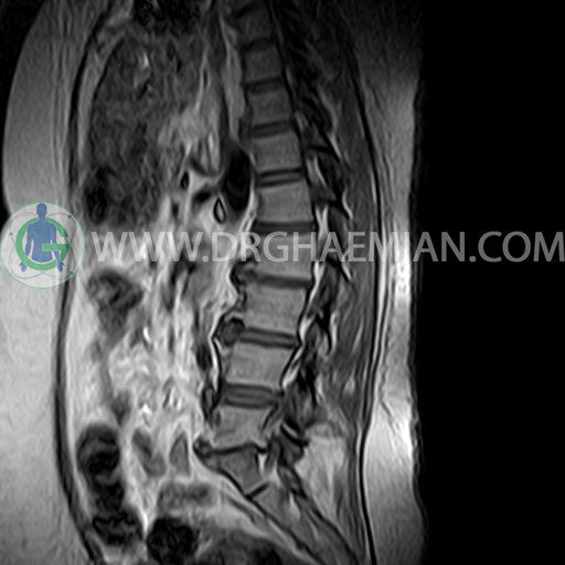
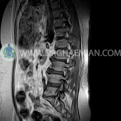
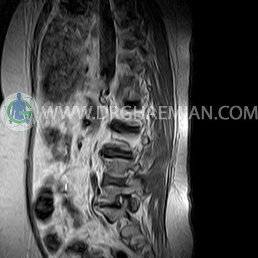
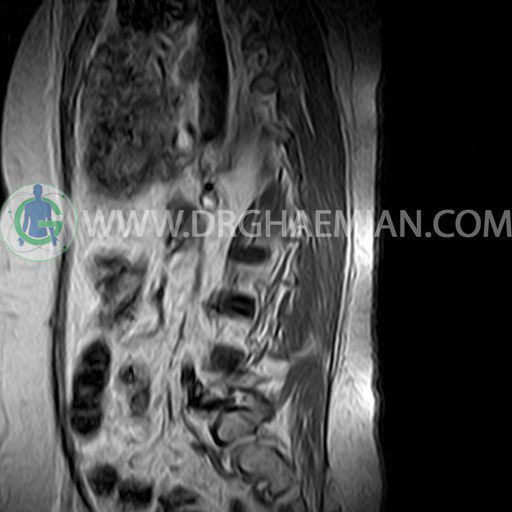
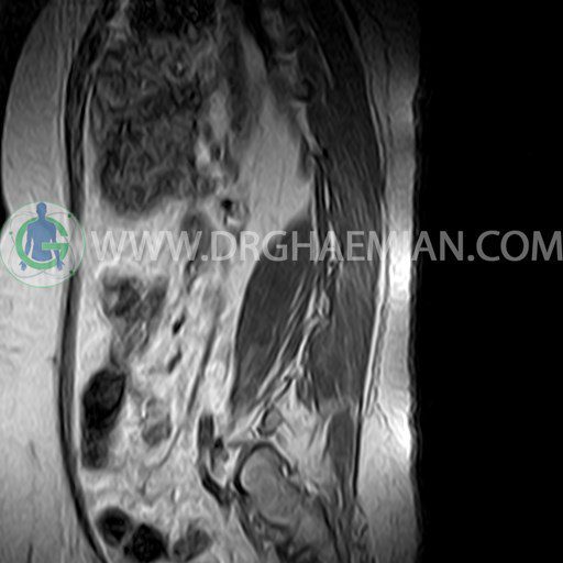
ام آر آی کمر از طریق انرژی آهنربا های قوی تصاویری از قسمت پایین ستون فقرات ( گودی کمر) ایجاد می کند. در این کیس استئوکندروز بین مهره ای کمر در دیسک L5/S1، کیفوسکولیوز، اسپوندیلوز، تنگ شدن فضای دیسک، دیهیدراته و بیرون زدگی دیسک های L1/L2 و L2/L3 و L3/L4 و L4/L5 دیده می شود.

گزارش پزشک :
LUMBOSACRAL SPINE MRI
(Without contrast)
Technique : Sagittal T1 , T2 , Axial T2 .
REPORT:
The imaged soft tissues show no abnormalities .
The visualized cord and filum terminalis are normal .
The conus medullaris terminates normally at L1.
Paravertebral stripe is normal in shape and signal intensity .
– Thoracolumbar kyphoscoliosis
– L1/L2 spondylosis, disc space narrowing, dehydration, bulging and right central protrusion with mild canal compromise
– L2/L3 spondylosis, disc space narrowing, dehydration, bulging and central annular fissure
– L3/L4 spondylosis, disc space narrowing, dehydration and bulging with canal – foramina stenosis
– L4/L5 disc dehydration, bulging and left central annular fissure with canal stenosis
– L5/S1 retrolisthesis grade 2 & intervertebral osteochondrosis
are seen.
