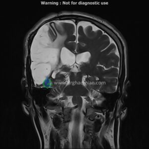
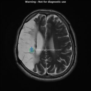
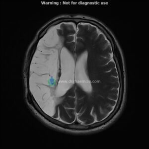
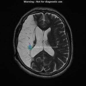
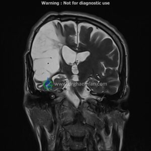
ام آر آی مغز یک روش تصویربرداری است که با استفاده از آهنربا های قوی و امواج رادیویی تصاویری از مغز و بافت های عصبی پیرامونی آن ایجاد می کند. در این کیس سکته مغزی و جراحی دیده میشود….

گزارش پزشک
BRAIN MRI
(Without contrast)
siemens MRI ( magnetom altea 1.5 tesla )
Technique:Axial & coronal FLAIR , Axial , sagittal , FSE T2 , coronal T1 .
REPORT :
The interhemispheric fissure is centered on the midline .
No abnormalities are seen in the basal ganglia , int . capsule, corpus callosum , Thalamus and tectal plate .
The brain stem , cerebellum show no abnormal changes in signal characteristics.
The sella and pituitary – pineal g .are normal and parasellar, suprasellar structures are unremarkable.
The cerebello pontine angle area appears normal on each side.
The internal acoustic meatus has normal width .
The orbital contents , falx are unremarkable .
Major intracranial vascular structures , pericavernous spaces and visualized intracranial nerve complex are normal .
– Diffuse cystic encephalomalacia in right MCA territory with gliosis and dilatation of right lateral ventricle suggestive for old infarction in right MCA territory
– Retention cyst in maxillary sinuses
– Right craniotomy with post-op changes
are seen.